Manganese »
PDB 5ekw-5fxv »
5f3o »
Manganese in PDB 5f3o: Crystal Structure of EHRNASEIII229 From Entamoeba Histolytica Complexed with MN2+
Protein crystallography data
The structure of Crystal Structure of EHRNASEIII229 From Entamoeba Histolytica Complexed with MN2+, PDB code: 5f3o
was solved by
X.Yu,
J.H.Gan,
J.B.Ma,
with X-Ray Crystallography technique. A brief refinement statistics is given in the table below:
| Resolution Low / High (Å) | 44.86 / 2.05 |
| Space group | P 21 21 21 |
| Cell size a, b, c (Å), α, β, γ (°) | 46.329, 89.729, 100.196, 90.00, 90.00, 90.00 |
| R / Rfree (%) | 19.7 / 24 |
Manganese Binding Sites:
The binding sites of Manganese atom in the Crystal Structure of EHRNASEIII229 From Entamoeba Histolytica Complexed with MN2+
(pdb code 5f3o). This binding sites where shown within
5.0 Angstroms radius around Manganese atom.
In total 2 binding sites of Manganese where determined in the Crystal Structure of EHRNASEIII229 From Entamoeba Histolytica Complexed with MN2+, PDB code: 5f3o:
Jump to Manganese binding site number: 1; 2;
In total 2 binding sites of Manganese where determined in the Crystal Structure of EHRNASEIII229 From Entamoeba Histolytica Complexed with MN2+, PDB code: 5f3o:
Jump to Manganese binding site number: 1; 2;
Manganese binding site 1 out of 2 in 5f3o
Go back to
Manganese binding site 1 out
of 2 in the Crystal Structure of EHRNASEIII229 From Entamoeba Histolytica Complexed with MN2+
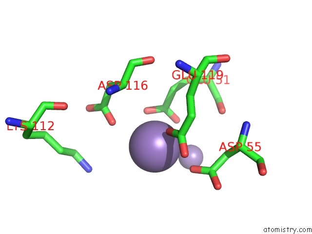
Mono view
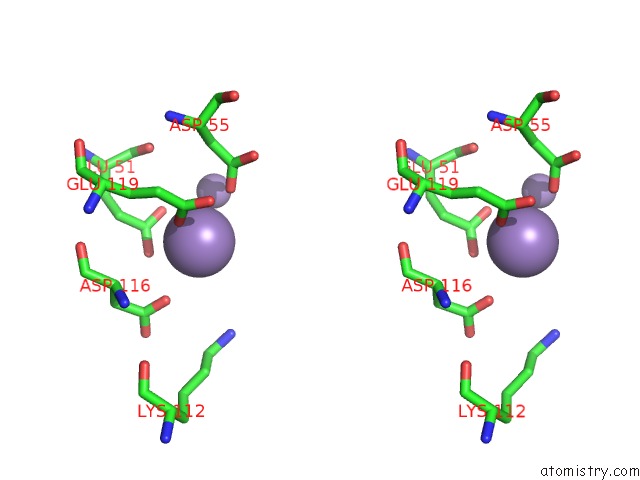
Stereo pair view
Mono view
Stereo pair view
A full contact list of Manganese with other atoms in the Mn binding
site number 1 of Crystal Structure of EHRNASEIII229 From Entamoeba Histolytica Complexed with MN2+ within 5.0Å range:
|
Manganese binding site 2 out of 2 in 5f3o
Go back to
Manganese binding site 2 out
of 2 in the Crystal Structure of EHRNASEIII229 From Entamoeba Histolytica Complexed with MN2+

Mono view
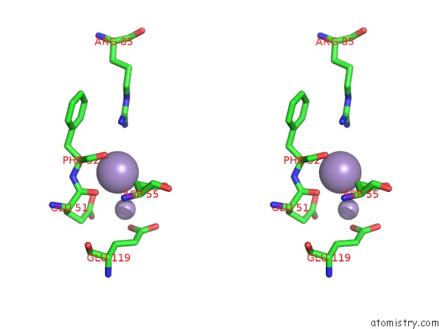
Stereo pair view
Mono view
Stereo pair view
A full contact list of Manganese with other atoms in the Mn binding
site number 2 of Crystal Structure of EHRNASEIII229 From Entamoeba Histolytica Complexed with MN2+ within 5.0Å range:
|
Reference:
X.Yu,
J.H.Gan,
J.B.Ma.
Structural and Functional Study of A Noncanonical Dicer Protein in Entamoeba Histolytica To Be Published.
Page generated: Sun Oct 6 00:10:50 2024
Last articles
K in 3CCLK in 3CCJ
K in 3CCE
K in 3CC7
K in 3CC4
K in 3CC2
K in 3C8S
K in 3C7R
K in 3C5D
K in 3C8Q