Manganese »
PDB 5ekw-5fxv »
5f1m »
Manganese in PDB 5f1m: Crystal Structure of Serine/Threonine Phosphatase STP1 From Staphylococcus Aureus
Enzymatic activity of Crystal Structure of Serine/Threonine Phosphatase STP1 From Staphylococcus Aureus
All present enzymatic activity of Crystal Structure of Serine/Threonine Phosphatase STP1 From Staphylococcus Aureus:
3.1.3.16;
3.1.3.16;
Protein crystallography data
The structure of Crystal Structure of Serine/Threonine Phosphatase STP1 From Staphylococcus Aureus, PDB code: 5f1m
was solved by
W.H.Zheng,
T.Wang,
Z.G.Li,
with X-Ray Crystallography technique. A brief refinement statistics is given in the table below:
| Resolution Low / High (Å) | 32.11 / 2.32 |
| Space group | P 2 21 21 |
| Cell size a, b, c (Å), α, β, γ (°) | 38.770, 77.350, 85.360, 90.00, 90.00, 90.00 |
| R / Rfree (%) | 20.2 / 25.6 |
Manganese Binding Sites:
The binding sites of Manganese atom in the Crystal Structure of Serine/Threonine Phosphatase STP1 From Staphylococcus Aureus
(pdb code 5f1m). This binding sites where shown within
5.0 Angstroms radius around Manganese atom.
In total 4 binding sites of Manganese where determined in the Crystal Structure of Serine/Threonine Phosphatase STP1 From Staphylococcus Aureus, PDB code: 5f1m:
Jump to Manganese binding site number: 1; 2; 3; 4;
In total 4 binding sites of Manganese where determined in the Crystal Structure of Serine/Threonine Phosphatase STP1 From Staphylococcus Aureus, PDB code: 5f1m:
Jump to Manganese binding site number: 1; 2; 3; 4;
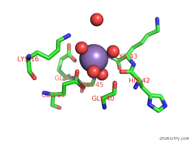
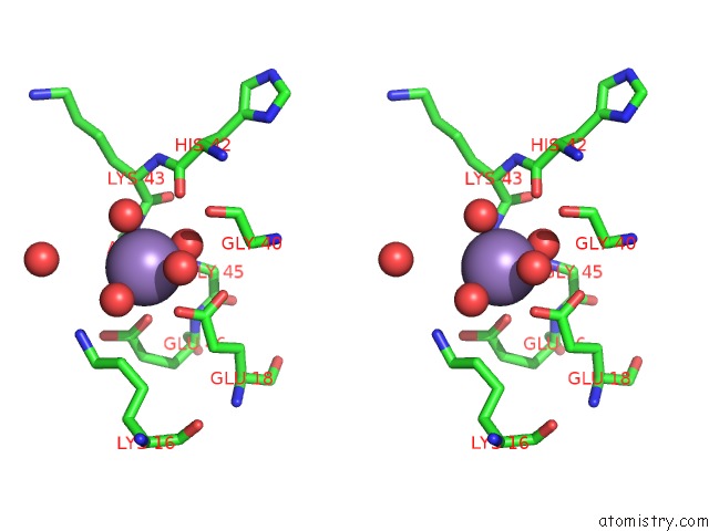
Manganese binding site 1 out of 4 in 5f1m
Go back to
Manganese binding site 1 out
of 4 in the Crystal Structure of Serine/Threonine Phosphatase STP1 From Staphylococcus Aureus
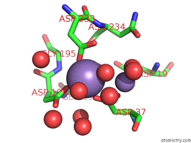
Mono view
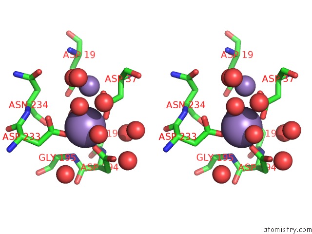
Stereo pair view
Mono view
Stereo pair view
A full contact list of Manganese with other atoms in the Mn binding
site number 1 of Crystal Structure of Serine/Threonine Phosphatase STP1 From Staphylococcus Aureus within 5.0Å range:
|
Manganese binding site 2 out of 4 in 5f1m
Go back to
Manganese binding site 2 out
of 4 in the Crystal Structure of Serine/Threonine Phosphatase STP1 From Staphylococcus Aureus
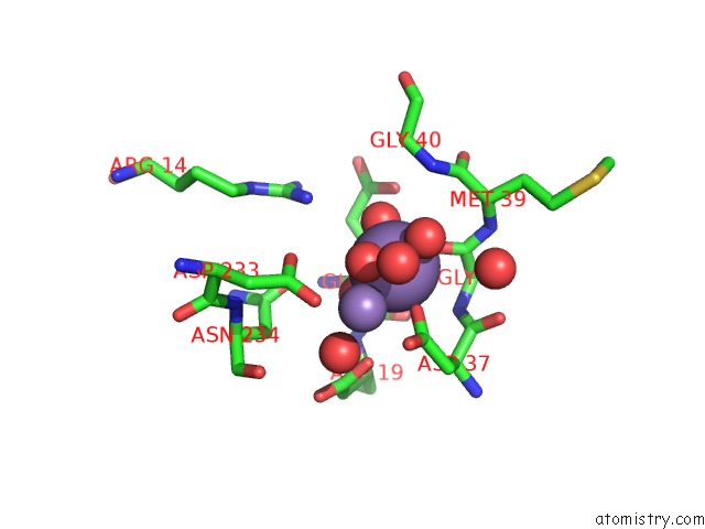
Mono view
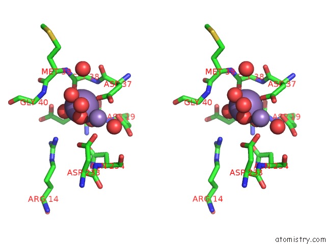
Stereo pair view
Mono view
Stereo pair view
A full contact list of Manganese with other atoms in the Mn binding
site number 2 of Crystal Structure of Serine/Threonine Phosphatase STP1 From Staphylococcus Aureus within 5.0Å range:
|
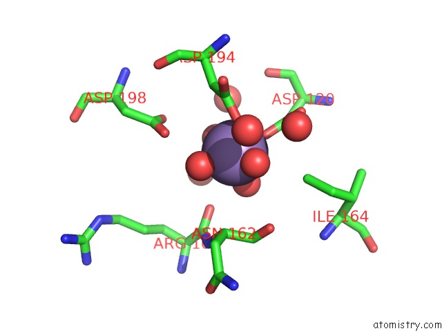
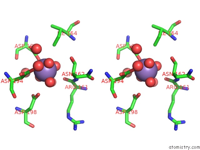
Manganese binding site 3 out of 4 in 5f1m
Go back to
Manganese binding site 3 out
of 4 in the Crystal Structure of Serine/Threonine Phosphatase STP1 From Staphylococcus Aureus
Mono view
Stereo pair view
Mono view
Stereo pair view
A full contact list of Manganese with other atoms in the Mn binding
site number 3 of Crystal Structure of Serine/Threonine Phosphatase STP1 From Staphylococcus Aureus within 5.0Å range:
|
Manganese binding site 4 out of 4 in 5f1m
Go back to
Manganese binding site 4 out
of 4 in the Crystal Structure of Serine/Threonine Phosphatase STP1 From Staphylococcus Aureus
Mono view
Stereo pair view
Mono view
Stereo pair view
A full contact list of Manganese with other atoms in the Mn binding
site number 4 of Crystal Structure of Serine/Threonine Phosphatase STP1 From Staphylococcus Aureus within 5.0Å range:
|
Reference:
W.Zheng,
X.Cai,
M.Xie,
Y.Liang,
T.Wang,
Z.Li.
Structure-Based Identification of A Potent Inhibitor Targeting STP1-Mediated Virulence Regulation in Staphylococcus Aureus Cell Chem Biol V. 23 1002 2016.
ISSN: ESSN 2451-9456
PubMed: 27499528
DOI: 10.1016/J.CHEMBIOL.2016.06.014
Page generated: Sun Oct 6 00:10:36 2024
ISSN: ESSN 2451-9456
PubMed: 27499528
DOI: 10.1016/J.CHEMBIOL.2016.06.014
Last articles
K in 1LW9K in 1LVG
K in 1LQP
K in 1LQK
K in 1LK0
K in 1LJU
K in 1LJL
K in 1LI9
K in 1L8I
K in 1LI0