Manganese »
PDB 5ekw-5fxv »
5esd »
Manganese in PDB 5esd: Crystal Structure of M. Tuberculosis Mend Bound to Thdp and MN2+
Enzymatic activity of Crystal Structure of M. Tuberculosis Mend Bound to Thdp and MN2+
All present enzymatic activity of Crystal Structure of M. Tuberculosis Mend Bound to Thdp and MN2+:
2.2.1.9;
2.2.1.9;
Protein crystallography data
The structure of Crystal Structure of M. Tuberculosis Mend Bound to Thdp and MN2+, PDB code: 5esd
was solved by
J.M.Johnston,
E.N.M.Jirgis,
G.Bashiri,
E.M.M.Bulloch,
E.N.Baker,
with X-Ray Crystallography technique. A brief refinement statistics is given in the table below:
| Resolution Low / High (Å) | 19.81 / 2.25 |
| Space group | P 21 21 21 |
| Cell size a, b, c (Å), α, β, γ (°) | 98.625, 138.657, 165.386, 90.00, 90.00, 90.00 |
| R / Rfree (%) | 21.1 / 23.5 |
Manganese Binding Sites:
The binding sites of Manganese atom in the Crystal Structure of M. Tuberculosis Mend Bound to Thdp and MN2+
(pdb code 5esd). This binding sites where shown within
5.0 Angstroms radius around Manganese atom.
In total 4 binding sites of Manganese where determined in the Crystal Structure of M. Tuberculosis Mend Bound to Thdp and MN2+, PDB code: 5esd:
Jump to Manganese binding site number: 1; 2; 3; 4;
In total 4 binding sites of Manganese where determined in the Crystal Structure of M. Tuberculosis Mend Bound to Thdp and MN2+, PDB code: 5esd:
Jump to Manganese binding site number: 1; 2; 3; 4;
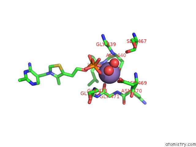
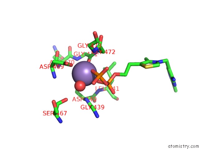
Manganese binding site 1 out of 4 in 5esd
Go back to
Manganese binding site 1 out
of 4 in the Crystal Structure of M. Tuberculosis Mend Bound to Thdp and MN2+
Mono view
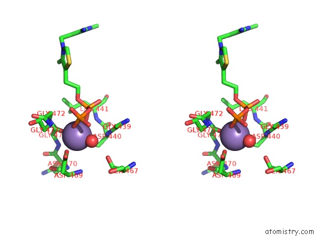
Stereo pair view
Mono view
Stereo pair view
A full contact list of Manganese with other atoms in the Mn binding
site number 1 of Crystal Structure of M. Tuberculosis Mend Bound to Thdp and MN2+ within 5.0Å range:
|
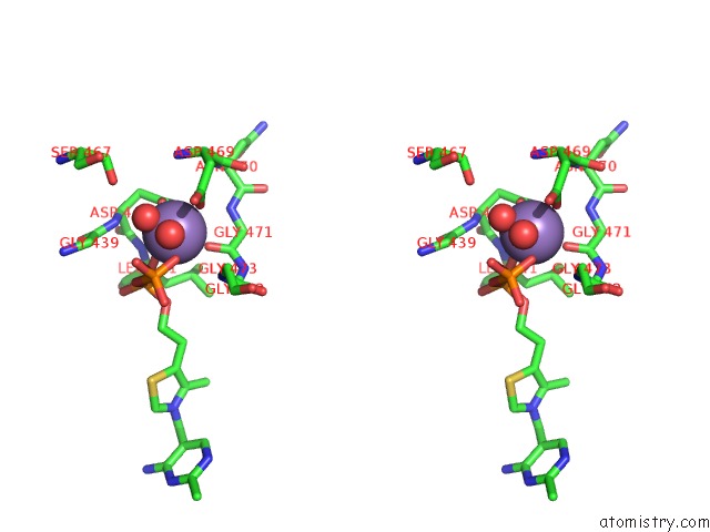
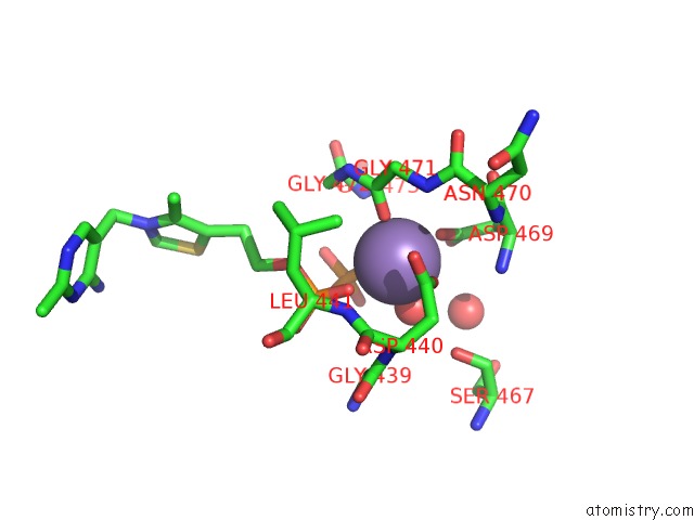
Manganese binding site 2 out of 4 in 5esd
Go back to
Manganese binding site 2 out
of 4 in the Crystal Structure of M. Tuberculosis Mend Bound to Thdp and MN2+
Mono view
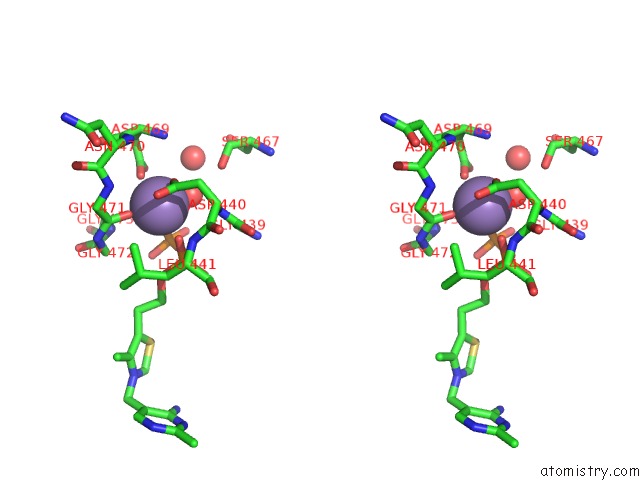
Stereo pair view
Mono view
Stereo pair view
A full contact list of Manganese with other atoms in the Mn binding
site number 2 of Crystal Structure of M. Tuberculosis Mend Bound to Thdp and MN2+ within 5.0Å range:
|
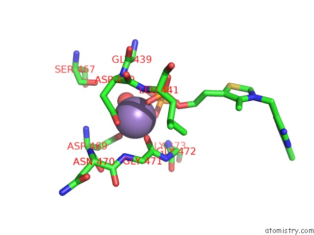
Manganese binding site 3 out of 4 in 5esd
Go back to
Manganese binding site 3 out
of 4 in the Crystal Structure of M. Tuberculosis Mend Bound to Thdp and MN2+
Mono view
Stereo pair view
Mono view
Stereo pair view
A full contact list of Manganese with other atoms in the Mn binding
site number 3 of Crystal Structure of M. Tuberculosis Mend Bound to Thdp and MN2+ within 5.0Å range:
|
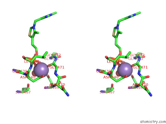
Manganese binding site 4 out of 4 in 5esd
Go back to
Manganese binding site 4 out
of 4 in the Crystal Structure of M. Tuberculosis Mend Bound to Thdp and MN2+
Mono view
Stereo pair view
Mono view
Stereo pair view
A full contact list of Manganese with other atoms in the Mn binding
site number 4 of Crystal Structure of M. Tuberculosis Mend Bound to Thdp and MN2+ within 5.0Å range:
|
Reference:
E.N.Jirgis,
G.Bashiri,
E.M.Bulloch,
J.M.Johnston,
E.N.Baker.
Structural Views Along the Mycobacterium Tuberculosis Mend Reaction Pathway Illuminate Key Aspects of Thiamin Diphosphate-Dependent Enzyme Mechanisms. Structure V. 24 1167 2016.
ISSN: ISSN 0969-2126
PubMed: 27291649
DOI: 10.1016/J.STR.2016.04.018
Page generated: Sun Oct 6 00:09:19 2024
ISSN: ISSN 0969-2126
PubMed: 27291649
DOI: 10.1016/J.STR.2016.04.018
Last articles
K in 3EUIK in 3EUM
K in 3EW9
K in 3EW8
K in 3EU8
K in 3ET8
K in 3ES0
K in 3EOG
K in 3EOH
K in 3ERU