Manganese »
PDB 4z8b-5a56 »
5a07 »
Manganese in PDB 5a07: X-Ray Structure of the Mannosyltransferase KTR4P From S. Cerevisiae in Complex with Gdp
Protein crystallography data
The structure of X-Ray Structure of the Mannosyltransferase KTR4P From S. Cerevisiae in Complex with Gdp, PDB code: 5a07
was solved by
D.D.D.Possner,
J.E.Guy,
with X-Ray Crystallography technique. A brief refinement statistics is given in the table below:
| Resolution Low / High (Å) | 86.79 / 1.90 |
| Space group | P 21 21 21 |
| Cell size a, b, c (Å), α, β, γ (°) | 61.215, 102.621, 162.651, 90.00, 90.00, 90.00 |
| R / Rfree (%) | 15.627 / 19.132 |
Manganese Binding Sites:
The binding sites of Manganese atom in the X-Ray Structure of the Mannosyltransferase KTR4P From S. Cerevisiae in Complex with Gdp
(pdb code 5a07). This binding sites where shown within
5.0 Angstroms radius around Manganese atom.
In total 2 binding sites of Manganese where determined in the X-Ray Structure of the Mannosyltransferase KTR4P From S. Cerevisiae in Complex with Gdp, PDB code: 5a07:
Jump to Manganese binding site number: 1; 2;
In total 2 binding sites of Manganese where determined in the X-Ray Structure of the Mannosyltransferase KTR4P From S. Cerevisiae in Complex with Gdp, PDB code: 5a07:
Jump to Manganese binding site number: 1; 2;
Manganese binding site 1 out of 2 in 5a07
Go back to
Manganese binding site 1 out
of 2 in the X-Ray Structure of the Mannosyltransferase KTR4P From S. Cerevisiae in Complex with Gdp
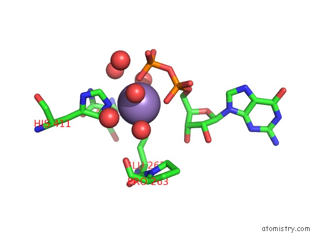
Mono view
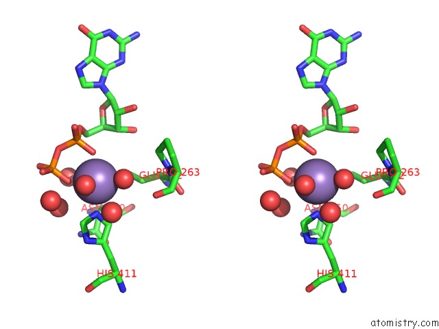
Stereo pair view
Mono view
Stereo pair view
A full contact list of Manganese with other atoms in the Mn binding
site number 1 of X-Ray Structure of the Mannosyltransferase KTR4P From S. Cerevisiae in Complex with Gdp within 5.0Å range:
|
Manganese binding site 2 out of 2 in 5a07
Go back to
Manganese binding site 2 out
of 2 in the X-Ray Structure of the Mannosyltransferase KTR4P From S. Cerevisiae in Complex with Gdp
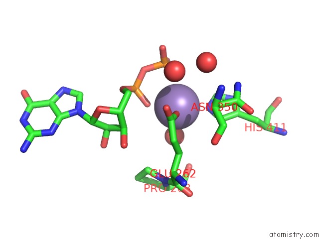
Mono view
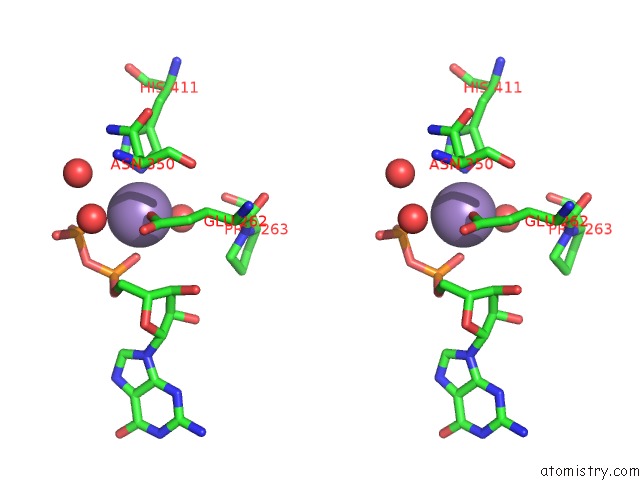
Stereo pair view
Mono view
Stereo pair view
A full contact list of Manganese with other atoms in the Mn binding
site number 2 of X-Ray Structure of the Mannosyltransferase KTR4P From S. Cerevisiae in Complex with Gdp within 5.0Å range:
|
Reference:
D.D.D.Possner,
M.Claesson,
J.E.Guy.
Structure of the Glycosyltransferase KTR4P From Saccharomyces Cerevisiae Plos One V. 10 36239 2015.
ISSN: ISSN 1932-6203
PubMed: 26296208
DOI: 10.1371/JOURNAL.PONE.013623
Page generated: Sat Oct 5 23:17:38 2024
ISSN: ISSN 1932-6203
PubMed: 26296208
DOI: 10.1371/JOURNAL.PONE.013623
Last articles
K in 5G0YK in 5G0X
K in 5G0I
K in 5FW3
K in 5G0J
K in 5G0R
K in 5FW2
K in 5G0H
K in 5FW1
K in 5FW5