Manganese »
PDB 4u87-4wiu »
4wie »
Manganese in PDB 4wie: Crystal Structure of Apo-Pepck From Mycobacterium Tuberculosis with Glycerol
Enzymatic activity of Crystal Structure of Apo-Pepck From Mycobacterium Tuberculosis with Glycerol
All present enzymatic activity of Crystal Structure of Apo-Pepck From Mycobacterium Tuberculosis with Glycerol:
4.1.1.32;
4.1.1.32;
Protein crystallography data
The structure of Crystal Structure of Apo-Pepck From Mycobacterium Tuberculosis with Glycerol, PDB code: 4wie
was solved by
H.L.Kim,
J.C.Sacchettini,
with X-Ray Crystallography technique. A brief refinement statistics is given in the table below:
| Resolution Low / High (Å) | 33.21 / 2.18 |
| Space group | C 1 2 1 |
| Cell size a, b, c (Å), α, β, γ (°) | 103.958, 122.564, 64.555, 90.00, 117.00, 90.00 |
| R / Rfree (%) | 13.8 / 19.1 |
Manganese Binding Sites:
The binding sites of Manganese atom in the Crystal Structure of Apo-Pepck From Mycobacterium Tuberculosis with Glycerol
(pdb code 4wie). This binding sites where shown within
5.0 Angstroms radius around Manganese atom.
In total only one binding site of Manganese was determined in the Crystal Structure of Apo-Pepck From Mycobacterium Tuberculosis with Glycerol, PDB code: 4wie:
In total only one binding site of Manganese was determined in the Crystal Structure of Apo-Pepck From Mycobacterium Tuberculosis with Glycerol, PDB code: 4wie:
Manganese binding site 1 out of 1 in 4wie
Go back to
Manganese binding site 1 out
of 1 in the Crystal Structure of Apo-Pepck From Mycobacterium Tuberculosis with Glycerol
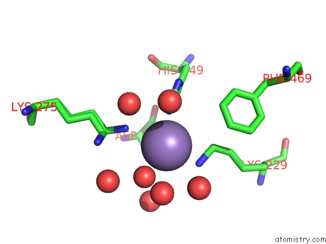
Mono view
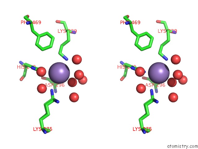
Stereo pair view
Mono view
Stereo pair view
A full contact list of Manganese with other atoms in the Mn binding
site number 1 of Crystal Structure of Apo-Pepck From Mycobacterium Tuberculosis with Glycerol within 5.0Å range:
|
Reference:
H.L.Kim,
J.C.Sacchettini.
Crystal Structure of Apo-Pepck From Mycobacterium Tuberculosis with Glycerol To Be Published.
Page generated: Sat Oct 5 21:34:03 2024
Last articles
Mg in 6XU1Mg in 6Y0Z
Mg in 6Y0T
Mg in 6Y0Y
Mg in 6Y09
Mg in 6XYD
Mg in 6XYB
Mg in 6XY5
Mg in 6XXC
Mg in 6XXQ