Manganese »
PDB 4qsh-4u3o »
4ruh »
Manganese in PDB 4ruh: Crystal Structure of Human Carnosinase-2 (CN2) in Complex with Inhibitor, Bestatin at 2.25 A
Enzymatic activity of Crystal Structure of Human Carnosinase-2 (CN2) in Complex with Inhibitor, Bestatin at 2.25 A
All present enzymatic activity of Crystal Structure of Human Carnosinase-2 (CN2) in Complex with Inhibitor, Bestatin at 2.25 A:
3.4.13.18;
3.4.13.18;
Protein crystallography data
The structure of Crystal Structure of Human Carnosinase-2 (CN2) in Complex with Inhibitor, Bestatin at 2.25 A, PDB code: 4ruh
was solved by
V.Pandya,
A.Kaushik,
A.K.Singh,
R.P.Singh,
S.Kumaran,
with X-Ray Crystallography technique. A brief refinement statistics is given in the table below:
| Resolution Low / High (Å) | 33.60 / 2.25 |
| Space group | P 21 21 21 |
| Cell size a, b, c (Å), α, β, γ (°) | 87.090, 100.070, 105.830, 90.00, 90.00, 90.00 |
| R / Rfree (%) | 21.9 / 28.2 |
Manganese Binding Sites:
The binding sites of Manganese atom in the Crystal Structure of Human Carnosinase-2 (CN2) in Complex with Inhibitor, Bestatin at 2.25 A
(pdb code 4ruh). This binding sites where shown within
5.0 Angstroms radius around Manganese atom.
In total 4 binding sites of Manganese where determined in the Crystal Structure of Human Carnosinase-2 (CN2) in Complex with Inhibitor, Bestatin at 2.25 A, PDB code: 4ruh:
Jump to Manganese binding site number: 1; 2; 3; 4;
In total 4 binding sites of Manganese where determined in the Crystal Structure of Human Carnosinase-2 (CN2) in Complex with Inhibitor, Bestatin at 2.25 A, PDB code: 4ruh:
Jump to Manganese binding site number: 1; 2; 3; 4;
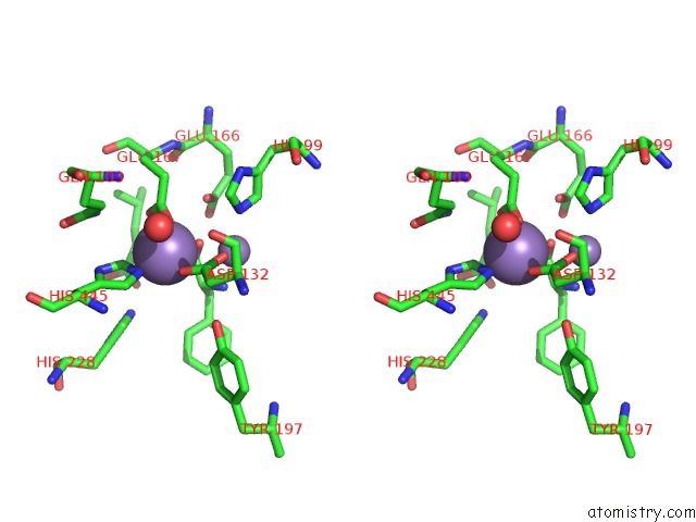
Manganese binding site 1 out of 4 in 4ruh
Go back to
Manganese binding site 1 out
of 4 in the Crystal Structure of Human Carnosinase-2 (CN2) in Complex with Inhibitor, Bestatin at 2.25 A
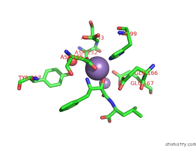
Mono view

Stereo pair view
Mono view
Stereo pair view
A full contact list of Manganese with other atoms in the Mn binding
site number 1 of Crystal Structure of Human Carnosinase-2 (CN2) in Complex with Inhibitor, Bestatin at 2.25 A within 5.0Å range:
|
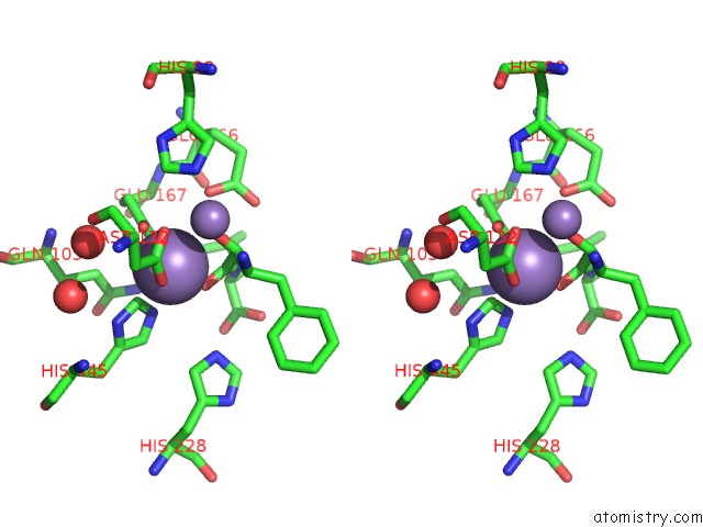
Manganese binding site 2 out of 4 in 4ruh
Go back to
Manganese binding site 2 out
of 4 in the Crystal Structure of Human Carnosinase-2 (CN2) in Complex with Inhibitor, Bestatin at 2.25 A
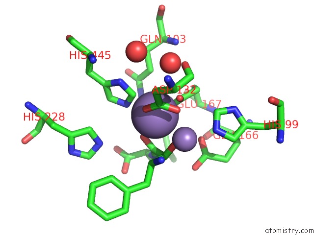
Mono view
Stereo pair view
Mono view
Stereo pair view
A full contact list of Manganese with other atoms in the Mn binding
site number 2 of Crystal Structure of Human Carnosinase-2 (CN2) in Complex with Inhibitor, Bestatin at 2.25 A within 5.0Å range:
|
Manganese binding site 3 out of 4 in 4ruh
Go back to
Manganese binding site 3 out
of 4 in the Crystal Structure of Human Carnosinase-2 (CN2) in Complex with Inhibitor, Bestatin at 2.25 A
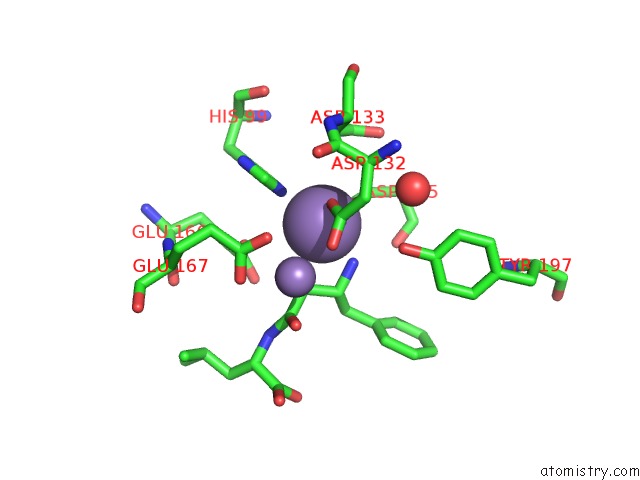
Mono view

Stereo pair view
Mono view
Stereo pair view
A full contact list of Manganese with other atoms in the Mn binding
site number 3 of Crystal Structure of Human Carnosinase-2 (CN2) in Complex with Inhibitor, Bestatin at 2.25 A within 5.0Å range:
|
Manganese binding site 4 out of 4 in 4ruh
Go back to
Manganese binding site 4 out
of 4 in the Crystal Structure of Human Carnosinase-2 (CN2) in Complex with Inhibitor, Bestatin at 2.25 A

Mono view
Stereo pair view
Mono view
Stereo pair view
A full contact list of Manganese with other atoms in the Mn binding
site number 4 of Crystal Structure of Human Carnosinase-2 (CN2) in Complex with Inhibitor, Bestatin at 2.25 A within 5.0Å range:
|
Reference:
V.Pandya,
A.Kaushik,
A.K.Singh,
R.P.Singh,
S.Kumaran.
Crystal Structure of Human Carnosinase-2 (CN2) in Complex with Inhibitor, Bestatin at 2.25 A To Be Published.
Page generated: Sat Oct 5 21:12:01 2024
Last articles
Mg in 2ZRWMg in 2ZRX
Mg in 2ZR8
Mg in 2ZRE
Mg in 2ZPI
Mg in 2ZPU
Mg in 2ZPG
Mg in 2ZPH
Mg in 2ZPE
Mg in 2ZPF