Manganese »
PDB 4qsh-4u3o »
4rhm »
Manganese in PDB 4rhm: Crystal Structure of T. Brucei Arginase-Like Protein Quadruple Mutant S149D/R151H/S153D/S226D
Enzymatic activity of Crystal Structure of T. Brucei Arginase-Like Protein Quadruple Mutant S149D/R151H/S153D/S226D
All present enzymatic activity of Crystal Structure of T. Brucei Arginase-Like Protein Quadruple Mutant S149D/R151H/S153D/S226D:
3.5.3.1;
3.5.3.1;
Protein crystallography data
The structure of Crystal Structure of T. Brucei Arginase-Like Protein Quadruple Mutant S149D/R151H/S153D/S226D, PDB code: 4rhm
was solved by
Y.Hai,
M.P.Barrett,
D.W.Christianson,
with X-Ray Crystallography technique. A brief refinement statistics is given in the table below:
| Resolution Low / High (Å) | 42.88 / 1.95 |
| Space group | C 1 2 1 |
| Cell size a, b, c (Å), α, β, γ (°) | 82.123, 137.350, 87.778, 90.00, 102.28, 90.00 |
| R / Rfree (%) | 20.1 / 23.7 |
Manganese Binding Sites:
The binding sites of Manganese atom in the Crystal Structure of T. Brucei Arginase-Like Protein Quadruple Mutant S149D/R151H/S153D/S226D
(pdb code 4rhm). This binding sites where shown within
5.0 Angstroms radius around Manganese atom.
In total 3 binding sites of Manganese where determined in the Crystal Structure of T. Brucei Arginase-Like Protein Quadruple Mutant S149D/R151H/S153D/S226D, PDB code: 4rhm:
Jump to Manganese binding site number: 1; 2; 3;
In total 3 binding sites of Manganese where determined in the Crystal Structure of T. Brucei Arginase-Like Protein Quadruple Mutant S149D/R151H/S153D/S226D, PDB code: 4rhm:
Jump to Manganese binding site number: 1; 2; 3;
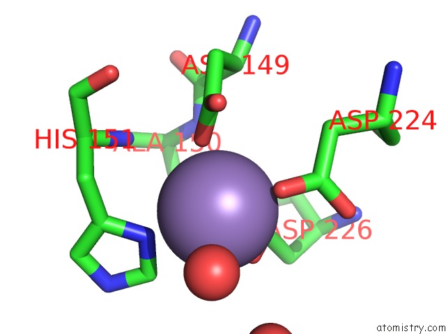
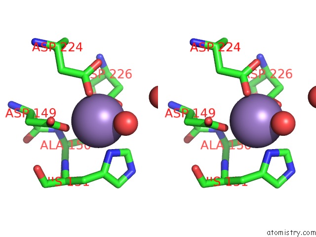
Manganese binding site 1 out of 3 in 4rhm
Go back to
Manganese binding site 1 out
of 3 in the Crystal Structure of T. Brucei Arginase-Like Protein Quadruple Mutant S149D/R151H/S153D/S226D
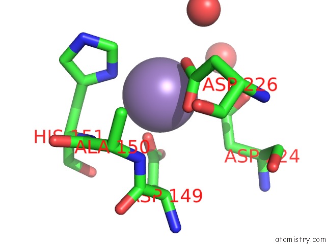
Mono view
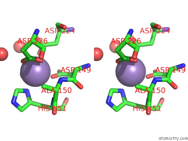
Stereo pair view
Mono view
Stereo pair view
A full contact list of Manganese with other atoms in the Mn binding
site number 1 of Crystal Structure of T. Brucei Arginase-Like Protein Quadruple Mutant S149D/R151H/S153D/S226D within 5.0Å range:
|
Manganese binding site 2 out of 3 in 4rhm
Go back to
Manganese binding site 2 out
of 3 in the Crystal Structure of T. Brucei Arginase-Like Protein Quadruple Mutant S149D/R151H/S153D/S226D
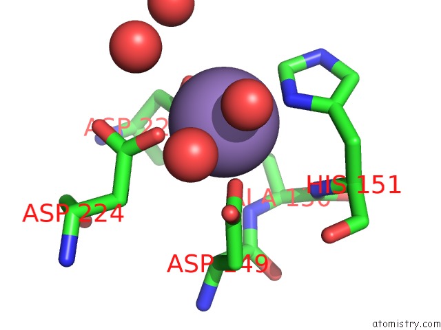
Mono view
Stereo pair view
Mono view
Stereo pair view
A full contact list of Manganese with other atoms in the Mn binding
site number 2 of Crystal Structure of T. Brucei Arginase-Like Protein Quadruple Mutant S149D/R151H/S153D/S226D within 5.0Å range:
|
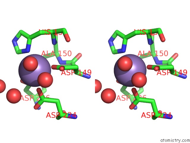
Manganese binding site 3 out of 3 in 4rhm
Go back to
Manganese binding site 3 out
of 3 in the Crystal Structure of T. Brucei Arginase-Like Protein Quadruple Mutant S149D/R151H/S153D/S226D
Mono view
Stereo pair view
Mono view
Stereo pair view
A full contact list of Manganese with other atoms in the Mn binding
site number 3 of Crystal Structure of T. Brucei Arginase-Like Protein Quadruple Mutant S149D/R151H/S153D/S226D within 5.0Å range:
|
Reference:
Y.Hai,
E.J.Kerkhoven,
M.P.Barrett,
D.W.Christianson.
Crystal Structure of An Arginase-Like Protein From Trypanosoma Brucei That Evolved Without A Binuclear Manganese Cluster. Biochemistry V. 54 458 2015.
ISSN: ISSN 0006-2960
PubMed: 25536859
DOI: 10.1021/BI501366A
Page generated: Sat Oct 5 21:11:14 2024
ISSN: ISSN 0006-2960
PubMed: 25536859
DOI: 10.1021/BI501366A
Last articles
K in 6DOOK in 6DON
K in 6DOM
K in 6DOP
K in 6DOL
K in 6DOK
K in 6DOJ
K in 6DOI
K in 6DOH
K in 6DOG