Manganese »
PDB 4qsh-4u3o »
4r60 »
Manganese in PDB 4r60: Crystal Structure of Xaa-Pro Dipeptidase From Xanthomonas Campestris
Enzymatic activity of Crystal Structure of Xaa-Pro Dipeptidase From Xanthomonas Campestris
All present enzymatic activity of Crystal Structure of Xaa-Pro Dipeptidase From Xanthomonas Campestris:
3.4.13.9;
3.4.13.9;
Protein crystallography data
The structure of Crystal Structure of Xaa-Pro Dipeptidase From Xanthomonas Campestris, PDB code: 4r60
was solved by
A.Kumar,
B.Ghosh,
V.N.Are,
S.N.Jamdar,
R.D.Makde,
S.M.Sharma,
with X-Ray Crystallography technique. A brief refinement statistics is given in the table below:
| Resolution Low / High (Å) | 46.46 / 1.83 |
| Space group | P 21 21 21 |
| Cell size a, b, c (Å), α, β, γ (°) | 84.324, 105.515, 111.352, 90.00, 90.00, 90.00 |
| R / Rfree (%) | 17.9 / 20.6 |
Other elements in 4r60:
The structure of Crystal Structure of Xaa-Pro Dipeptidase From Xanthomonas Campestris also contains other interesting chemical elements:
| Sodium | (Na) | 2 atoms |
Manganese Binding Sites:
The binding sites of Manganese atom in the Crystal Structure of Xaa-Pro Dipeptidase From Xanthomonas Campestris
(pdb code 4r60). This binding sites where shown within
5.0 Angstroms radius around Manganese atom.
In total 4 binding sites of Manganese where determined in the Crystal Structure of Xaa-Pro Dipeptidase From Xanthomonas Campestris, PDB code: 4r60:
Jump to Manganese binding site number: 1; 2; 3; 4;
In total 4 binding sites of Manganese where determined in the Crystal Structure of Xaa-Pro Dipeptidase From Xanthomonas Campestris, PDB code: 4r60:
Jump to Manganese binding site number: 1; 2; 3; 4;
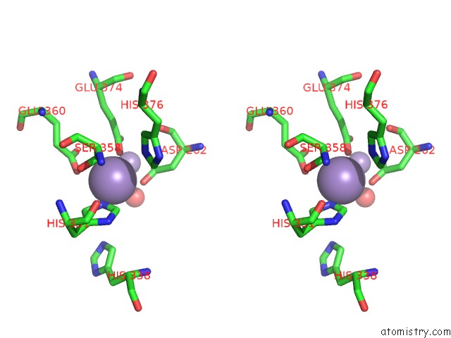
Manganese binding site 1 out of 4 in 4r60
Go back to
Manganese binding site 1 out
of 4 in the Crystal Structure of Xaa-Pro Dipeptidase From Xanthomonas Campestris
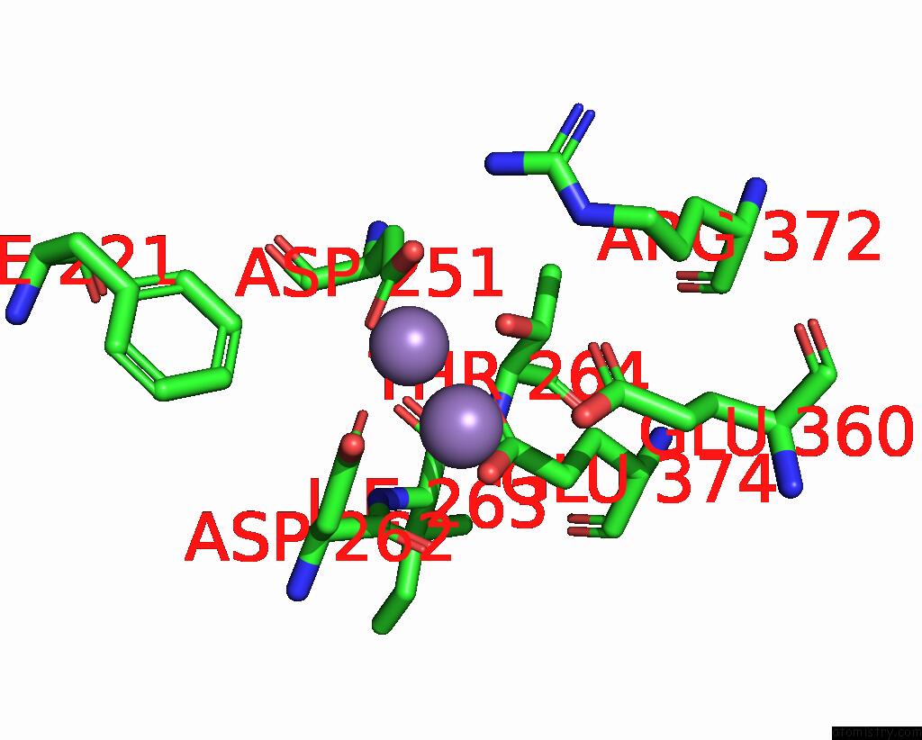
Mono view
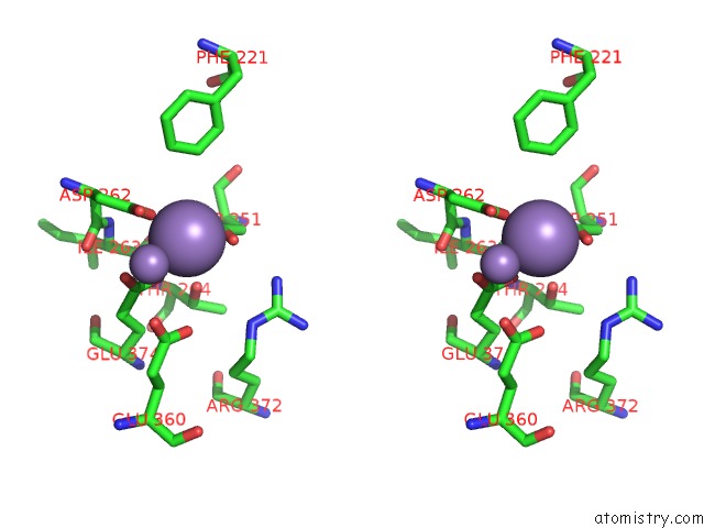
Stereo pair view
Mono view
Stereo pair view
A full contact list of Manganese with other atoms in the Mn binding
site number 1 of Crystal Structure of Xaa-Pro Dipeptidase From Xanthomonas Campestris within 5.0Å range:
|
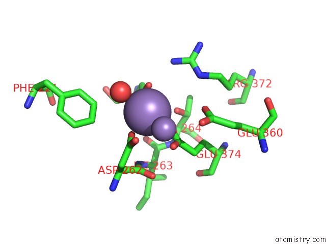
Manganese binding site 2 out of 4 in 4r60
Go back to
Manganese binding site 2 out
of 4 in the Crystal Structure of Xaa-Pro Dipeptidase From Xanthomonas Campestris
Mono view
Stereo pair view
Mono view
Stereo pair view
A full contact list of Manganese with other atoms in the Mn binding
site number 2 of Crystal Structure of Xaa-Pro Dipeptidase From Xanthomonas Campestris within 5.0Å range:
|
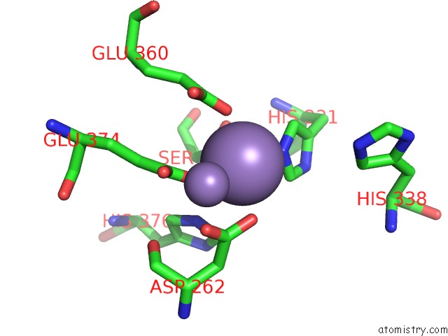
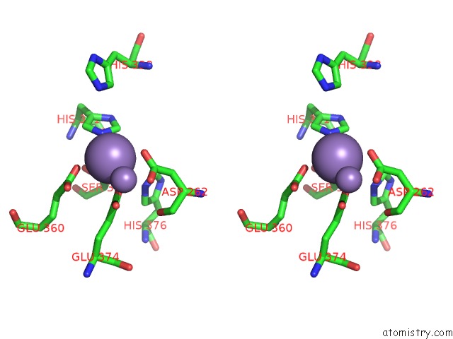
Manganese binding site 3 out of 4 in 4r60
Go back to
Manganese binding site 3 out
of 4 in the Crystal Structure of Xaa-Pro Dipeptidase From Xanthomonas Campestris

Mono view
Stereo pair view
Mono view
Stereo pair view
A full contact list of Manganese with other atoms in the Mn binding
site number 3 of Crystal Structure of Xaa-Pro Dipeptidase From Xanthomonas Campestris within 5.0Å range:
|
Manganese binding site 4 out of 4 in 4r60
Go back to
Manganese binding site 4 out
of 4 in the Crystal Structure of Xaa-Pro Dipeptidase From Xanthomonas Campestris
Mono view

Stereo pair view
Mono view
Stereo pair view
A full contact list of Manganese with other atoms in the Mn binding
site number 4 of Crystal Structure of Xaa-Pro Dipeptidase From Xanthomonas Campestris within 5.0Å range:
|
Reference:
A.Kumar,
B.Ghosh,
V.N.Are,
S.N.Jamdar,
R.D.Makde,
S.M.Sharma.
Crystal Structure of Xaa-Pro Dipeptidase From Xanthomonas Campestris To Be Published.
Page generated: Sat Oct 5 21:05:48 2024
Last articles
Mg in 3CF5Mg in 3CCV
Mg in 3CCU
Mg in 3CCS
Mg in 3CCR
Mg in 3CCQ
Mg in 3CCM
Mg in 3CCL
Mg in 3CCJ
Mg in 3CC2