Manganese »
PDB 4nxz-4phd »
4pcl »
Manganese in PDB 4pcl: X-Ray Crystal Structure of An O-Methyltransferase From Anaplasma Phagocytophilum Bound to Sam and A Manganese Ion.
Protein crystallography data
The structure of X-Ray Crystal Structure of An O-Methyltransferase From Anaplasma Phagocytophilum Bound to Sam and A Manganese Ion., PDB code: 4pcl
was solved by
J.W.Fairman,
T.E.Edwards,
D.Lorimer,
Seattle Structural Genomics Centerfor Infectious Disease (Ssgcid),
with X-Ray Crystallography technique. A brief refinement statistics is given in the table below:
| Resolution Low / High (Å) | 37.84 / 1.85 |
| Space group | P 63 2 2 |
| Cell size a, b, c (Å), α, β, γ (°) | 123.870, 123.870, 121.310, 90.00, 90.00, 120.00 |
| R / Rfree (%) | 17 / 20.9 |
Manganese Binding Sites:
The binding sites of Manganese atom in the X-Ray Crystal Structure of An O-Methyltransferase From Anaplasma Phagocytophilum Bound to Sam and A Manganese Ion.
(pdb code 4pcl). This binding sites where shown within
5.0 Angstroms radius around Manganese atom.
In total 2 binding sites of Manganese where determined in the X-Ray Crystal Structure of An O-Methyltransferase From Anaplasma Phagocytophilum Bound to Sam and A Manganese Ion., PDB code: 4pcl:
Jump to Manganese binding site number: 1; 2;
In total 2 binding sites of Manganese where determined in the X-Ray Crystal Structure of An O-Methyltransferase From Anaplasma Phagocytophilum Bound to Sam and A Manganese Ion., PDB code: 4pcl:
Jump to Manganese binding site number: 1; 2;
Manganese binding site 1 out of 2 in 4pcl
Go back to
Manganese binding site 1 out
of 2 in the X-Ray Crystal Structure of An O-Methyltransferase From Anaplasma Phagocytophilum Bound to Sam and A Manganese Ion.
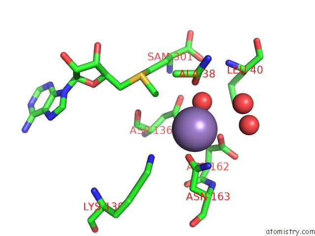
Mono view
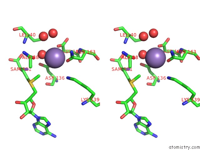
Stereo pair view
Mono view
Stereo pair view
A full contact list of Manganese with other atoms in the Mn binding
site number 1 of X-Ray Crystal Structure of An O-Methyltransferase From Anaplasma Phagocytophilum Bound to Sam and A Manganese Ion. within 5.0Å range:
|
Manganese binding site 2 out of 2 in 4pcl
Go back to
Manganese binding site 2 out
of 2 in the X-Ray Crystal Structure of An O-Methyltransferase From Anaplasma Phagocytophilum Bound to Sam and A Manganese Ion.
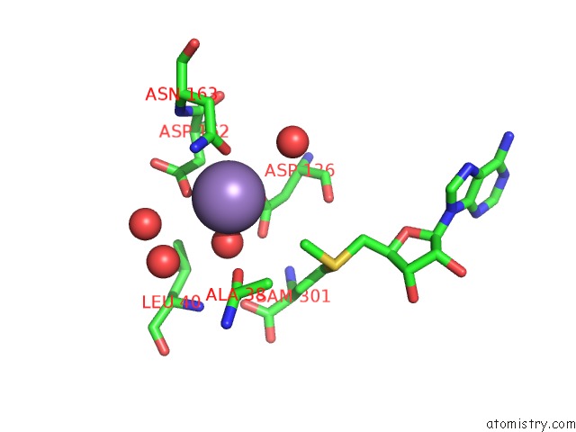
Mono view
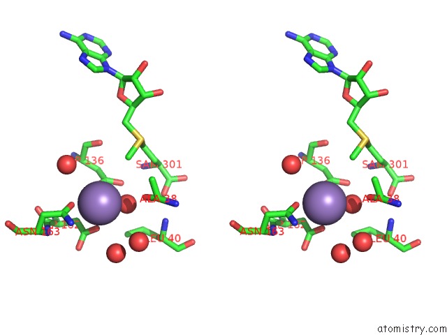
Stereo pair view
Mono view
Stereo pair view
A full contact list of Manganese with other atoms in the Mn binding
site number 2 of X-Ray Crystal Structure of An O-Methyltransferase From Anaplasma Phagocytophilum Bound to Sam and A Manganese Ion. within 5.0Å range:
|
Reference:
J.W.Fairman,
T.E.Edwards,
D.Lorimer.
X-Ray Crystal Structure of An O-Methyltransferase From Anaplasma Phagocytophilum Bound to Sam and A Manganese Ion. To Be Published.
Page generated: Sat Oct 5 20:45:02 2024
Last articles
Mg in 3DYMMg in 3E18
Mg in 3DZO
Mg in 3DZE
Mg in 3DYS
Mg in 3DYG
Mg in 3DYN
Mg in 3DYH
Mg in 3DYF
Mg in 3DYL