Manganese »
PDB 4nxz-4phd »
4pca »
Manganese in PDB 4pca: X-Ray Crystal Structure of An O-Methyltransferase From Anaplasma Phagocytophilum Bound to Sah and Manganese
Protein crystallography data
The structure of X-Ray Crystal Structure of An O-Methyltransferase From Anaplasma Phagocytophilum Bound to Sah and Manganese, PDB code: 4pca
was solved by
J.W.Fairman,
J.Abendroth,
D.Lorimer,
T.E.Edwards,
Seattle Structuralgenomics Center For Infectious Disease (Ssgcid),
with X-Ray Crystallography technique. A brief refinement statistics is given in the table below:
| Resolution Low / High (Å) | 40.58 / 1.50 |
| Space group | P 21 21 21 |
| Cell size a, b, c (Å), α, β, γ (°) | 85.210, 102.760, 103.320, 90.00, 90.00, 90.00 |
| R / Rfree (%) | 14.7 / 17.2 |
Manganese Binding Sites:
The binding sites of Manganese atom in the X-Ray Crystal Structure of An O-Methyltransferase From Anaplasma Phagocytophilum Bound to Sah and Manganese
(pdb code 4pca). This binding sites where shown within
5.0 Angstroms radius around Manganese atom.
In total 4 binding sites of Manganese where determined in the X-Ray Crystal Structure of An O-Methyltransferase From Anaplasma Phagocytophilum Bound to Sah and Manganese, PDB code: 4pca:
Jump to Manganese binding site number: 1; 2; 3; 4;
In total 4 binding sites of Manganese where determined in the X-Ray Crystal Structure of An O-Methyltransferase From Anaplasma Phagocytophilum Bound to Sah and Manganese, PDB code: 4pca:
Jump to Manganese binding site number: 1; 2; 3; 4;
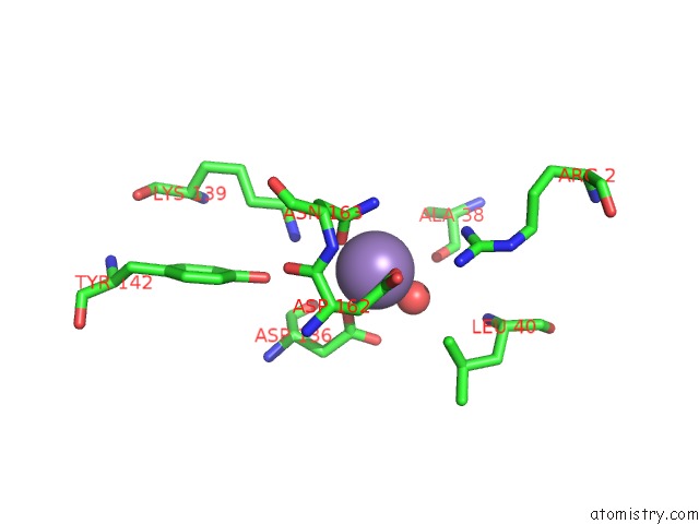
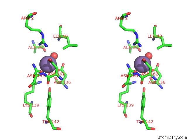
Manganese binding site 1 out of 4 in 4pca
Go back to
Manganese binding site 1 out
of 4 in the X-Ray Crystal Structure of An O-Methyltransferase From Anaplasma Phagocytophilum Bound to Sah and Manganese
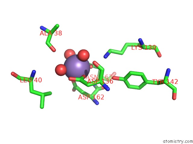
Mono view
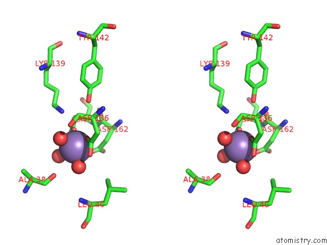
Stereo pair view
Mono view
Stereo pair view
A full contact list of Manganese with other atoms in the Mn binding
site number 1 of X-Ray Crystal Structure of An O-Methyltransferase From Anaplasma Phagocytophilum Bound to Sah and Manganese within 5.0Å range:
|
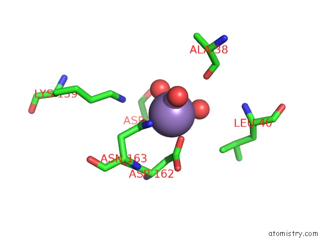
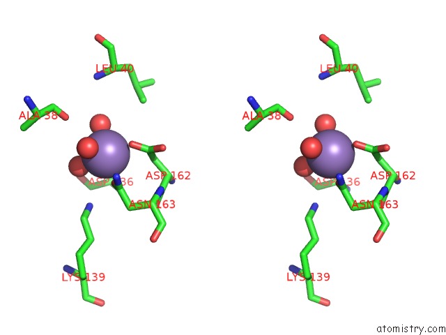
Manganese binding site 2 out of 4 in 4pca
Go back to
Manganese binding site 2 out
of 4 in the X-Ray Crystal Structure of An O-Methyltransferase From Anaplasma Phagocytophilum Bound to Sah and Manganese
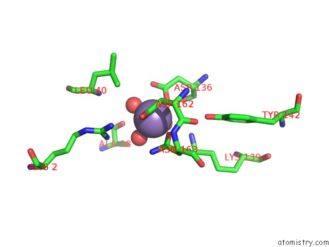
Mono view
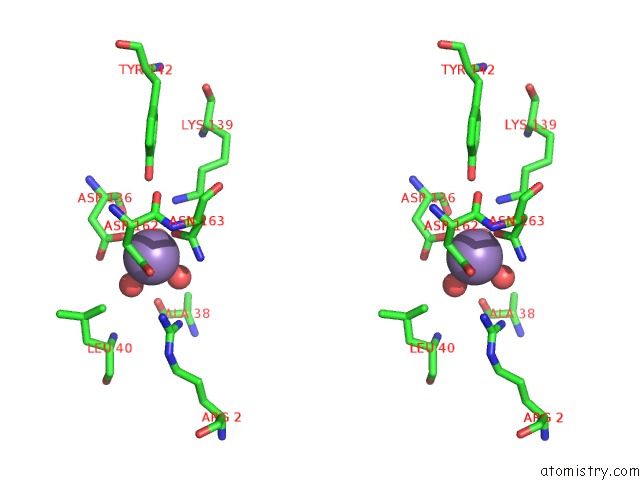
Stereo pair view
Mono view
Stereo pair view
A full contact list of Manganese with other atoms in the Mn binding
site number 2 of X-Ray Crystal Structure of An O-Methyltransferase From Anaplasma Phagocytophilum Bound to Sah and Manganese within 5.0Å range:
|
Manganese binding site 3 out of 4 in 4pca
Go back to
Manganese binding site 3 out
of 4 in the X-Ray Crystal Structure of An O-Methyltransferase From Anaplasma Phagocytophilum Bound to Sah and Manganese
Mono view
Stereo pair view
Mono view
Stereo pair view
A full contact list of Manganese with other atoms in the Mn binding
site number 3 of X-Ray Crystal Structure of An O-Methyltransferase From Anaplasma Phagocytophilum Bound to Sah and Manganese within 5.0Å range:
|
Manganese binding site 4 out of 4 in 4pca
Go back to
Manganese binding site 4 out
of 4 in the X-Ray Crystal Structure of An O-Methyltransferase From Anaplasma Phagocytophilum Bound to Sah and Manganese
Mono view
Stereo pair view
Mono view
Stereo pair view
A full contact list of Manganese with other atoms in the Mn binding
site number 4 of X-Ray Crystal Structure of An O-Methyltransferase From Anaplasma Phagocytophilum Bound to Sah and Manganese within 5.0Å range:
|
Reference:
J.W.Fairman,
J.Abendroth,
D.Lorimer,
T.E.Edwards.
X-Ray Crystal Structure of An O-Methyltransferase From Anaplasma Phagocytophilum Bound to Sah and Manganese To Be Published.
Page generated: Sat Oct 5 20:44:55 2024
Last articles
Mg in 3DY8Mg in 3DVL
Mg in 3DY7
Mg in 3DXJ
Mg in 3DV0
Mg in 3DV9
Mg in 3DV6
Mg in 3DV3
Mg in 3DV4
Mg in 3DUF