Manganese »
PDB 4nxz-4phd »
4ogc »
Manganese in PDB 4ogc: Crystal Structure of the Type II-C CAS9 Enzyme From Actinomyces Naeslundii
Protein crystallography data
The structure of Crystal Structure of the Type II-C CAS9 Enzyme From Actinomyces Naeslundii, PDB code: 4ogc
was solved by
F.Jiang,
E.Ma,
S.Lin,
J.A.Doudna,
with X-Ray Crystallography technique. A brief refinement statistics is given in the table below:
| Resolution Low / High (Å) | 68.30 / 2.80 |
| Space group | P 1 21 1 |
| Cell size a, b, c (Å), α, β, γ (°) | 74.610, 132.560, 80.040, 90.00, 95.38, 90.00 |
| R / Rfree (%) | 19.4 / 23.3 |
Other elements in 4ogc:
The structure of Crystal Structure of the Type II-C CAS9 Enzyme From Actinomyces Naeslundii also contains other interesting chemical elements:
| Magnesium | (Mg) | 2 atoms |
| Zinc | (Zn) | 1 atom |
Manganese Binding Sites:
The binding sites of Manganese atom in the Crystal Structure of the Type II-C CAS9 Enzyme From Actinomyces Naeslundii
(pdb code 4ogc). This binding sites where shown within
5.0 Angstroms radius around Manganese atom.
In total 2 binding sites of Manganese where determined in the Crystal Structure of the Type II-C CAS9 Enzyme From Actinomyces Naeslundii, PDB code: 4ogc:
Jump to Manganese binding site number: 1; 2;
In total 2 binding sites of Manganese where determined in the Crystal Structure of the Type II-C CAS9 Enzyme From Actinomyces Naeslundii, PDB code: 4ogc:
Jump to Manganese binding site number: 1; 2;
Manganese binding site 1 out of 2 in 4ogc
Go back to
Manganese binding site 1 out
of 2 in the Crystal Structure of the Type II-C CAS9 Enzyme From Actinomyces Naeslundii
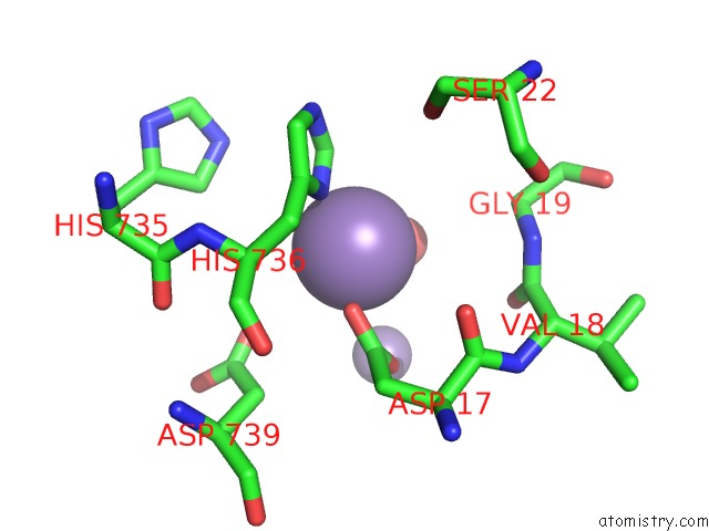
Mono view
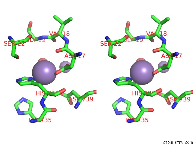
Stereo pair view
Mono view
Stereo pair view
A full contact list of Manganese with other atoms in the Mn binding
site number 1 of Crystal Structure of the Type II-C CAS9 Enzyme From Actinomyces Naeslundii within 5.0Å range:
|
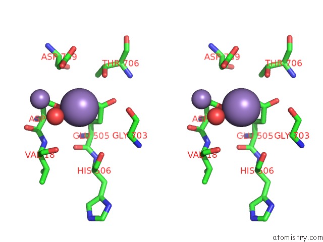
Manganese binding site 2 out of 2 in 4ogc
Go back to
Manganese binding site 2 out
of 2 in the Crystal Structure of the Type II-C CAS9 Enzyme From Actinomyces Naeslundii
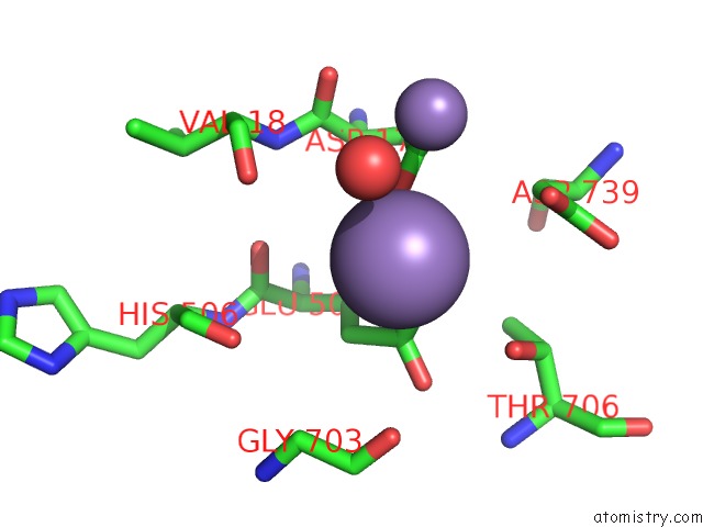
Mono view
Stereo pair view
Mono view
Stereo pair view
A full contact list of Manganese with other atoms in the Mn binding
site number 2 of Crystal Structure of the Type II-C CAS9 Enzyme From Actinomyces Naeslundii within 5.0Å range:
|
Reference:
M.Jinek,
F.Jiang,
D.W.Taylor,
S.H.Sternberg,
E.Kaya,
E.Ma,
C.Anders,
M.Hauer,
K.Zhou,
S.Lin,
M.Kaplan,
A.T.Iavarone,
E.Charpentier,
E.Nogales,
J.A.Doudna.
Structures of CAS9 Endonucleases Reveal Rna-Mediated Conformational Activation. Science V. 343 47997 2014.
ISSN: ISSN 0036-8075
PubMed: 24505130
DOI: 10.1126/SCIENCE.1247997
Page generated: Sat Oct 5 20:42:16 2024
ISSN: ISSN 0036-8075
PubMed: 24505130
DOI: 10.1126/SCIENCE.1247997
Last articles
K in 3POXK in 3Q8M
K in 3Q3V
K in 3Q8L
K in 3Q8K
K in 3PW3
K in 3Q8H
K in 3Q1Y
K in 3PIO
K in 3PY6