Atomistry » Manganese » PDB 4lta-4mu3 » 4mov
Atomistry »
  Manganese »
    PDB 4lta-4mu3 »
      4mov »

Manganese in PDB 4mov: 1.45 A Resolution Crystal Structure of Protein Phosphatase 1

Enzymatic activity of 1.45 A Resolution Crystal Structure of Protein Phosphatase 1

All present enzymatic activity of 1.45 A Resolution Crystal Structure of Protein Phosphatase 1:
3.1.3.16;

Protein crystallography data

The structure of 1.45 A Resolution Crystal Structure of Protein Phosphatase 1, PDB code: 4mov was solved by M.S.Choy, W.Peti, R.Page, with X-Ray Crystallography technique. A brief refinement statistics is given in the table below:

Resolution Low / High (Å) 46.93 / 1.45
Space group P 21 21 21
Cell size a, b, c (Å), α, β, γ (°) 65.724, 77.600, 133.035, 90.00, 90.00, 90.00
R / Rfree (%) 15 / 16.7

Other elements in 4mov:

The structure of 1.45 A Resolution Crystal Structure of Protein Phosphatase 1 also contains other interesting chemical elements:

Chlorine (Cl) 2 atoms

Manganese Binding Sites:

The binding sites of Manganese atom in the 1.45 A Resolution Crystal Structure of Protein Phosphatase 1 (pdb code 4mov). This binding sites where shown within 5.0 Angstroms radius around Manganese atom.
In total 4 binding sites of Manganese where determined in the 1.45 A Resolution Crystal Structure of Protein Phosphatase 1, PDB code: 4mov:
Jump to Manganese binding site number: 1; 2; 3; 4;

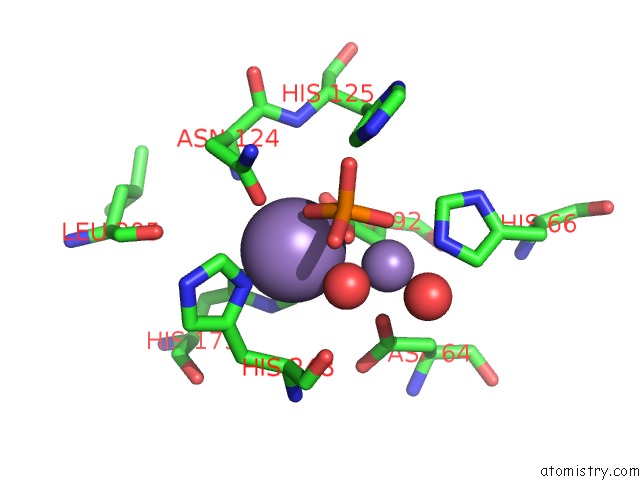
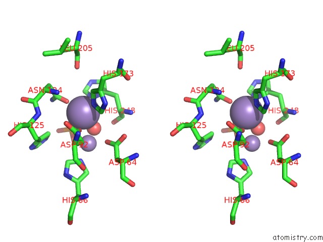
Manganese binding site 1 out of 4 in 4mov

Go back to Manganese Binding Sites List in 4mov
Manganese binding site 1 out of 4 in the 1.45 A Resolution Crystal Structure of Protein Phosphatase 1


Mono view


Stereo pair view

A full contact list of Manganese with other atoms in the Mn binding site number 1 of 1.45 A Resolution Crystal Structure of Protein Phosphatase 1 within 5.0Å range:
probe atom residue distance (Å) B Occ
A:Mn401

b:9.0
occ:1.00
OD1 A:ASN124 2.1 7.9 1.0
O3 A:PO4403 2.2 11.4 0.8
NE2 A:HIS173 2.2 7.7 1.0
OD2 A:ASP92 2.2 8.2 1.0
ND1 A:HIS248 2.2 10.6 1.0
O A:HOH737 2.3 13.0 1.0
CE1 A:HIS248 3.1 11.6 1.0
MN A:MN402 3.1 13.4 0.5
CG A:ASP92 3.2 7.8 1.0
CE1 A:HIS173 3.2 7.3 1.0
CG A:ASN124 3.2 9.2 1.0
CD2 A:HIS173 3.2 6.5 1.0
CG A:HIS248 3.4 9.0 1.0
P A:PO4403 3.4 17.4 0.8
OD1 A:ASP92 3.5 7.5 1.0
ND2 A:ASN124 3.6 10.9 1.0
O1 A:PO4403 3.7 19.6 0.8
CA A:HIS248 3.8 9.9 1.0
CB A:HIS248 3.9 9.5 1.0
OD2 A:ASP64 4.0 11.8 1.0
O4 A:PO4403 4.1 19.7 0.8
O A:HIS248 4.1 17.6 1.0
CD2 A:HIS125 4.2 9.3 1.0
NE2 A:HIS248 4.3 13.5 1.0
ND1 A:HIS173 4.3 6.9 1.0
CG A:HIS173 4.3 7.1 1.0
CD2 A:HIS248 4.4 14.3 1.0
CB A:ASP92 4.4 6.9 1.0
CB A:ASN124 4.5 8.6 1.0
C A:HIS248 4.5 12.0 1.0
N A:ASN124 4.5 7.5 1.0
O2 A:PO4403 4.6 18.8 0.8
O A:LEU205 4.7 10.1 1.0
NE2 A:HIS125 4.8 9.8 1.0
N A:HIS248 4.9 9.5 1.0
NE2 A:HIS66 4.9 9.6 1.0
O A:HOH746 4.9 33.5 1.0
CG A:ASP64 4.9 9.8 1.0

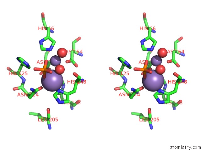
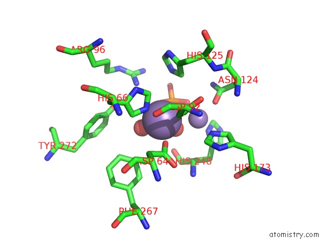
Manganese binding site 2 out of 4 in 4mov

Go back to Manganese Binding Sites List in 4mov
Manganese binding site 2 out of 4 in the 1.45 A Resolution Crystal Structure of Protein Phosphatase 1


Mono view


Stereo pair view

A full contact list of Manganese with other atoms in the Mn binding site number 2 of 1.45 A Resolution Crystal Structure of Protein Phosphatase 1 within 5.0Å range:
probe atom residue distance (Å) B Occ
A:Mn402

b:13.4
occ:0.50
O A:HOH737 1.7 13.0 1.0
NE2 A:HIS66 2.1 9.6 1.0
OD2 A:ASP92 2.2 8.2 1.0
O1 A:PO4403 2.2 19.6 0.8
O A:HOH746 2.3 33.5 1.0
OD2 A:ASP64 2.3 11.8 1.0
CE1 A:HIS66 3.0 8.4 1.0
CD2 A:HIS66 3.1 9.2 1.0
MN A:MN401 3.1 9.0 1.0
CG A:ASP92 3.3 7.8 1.0
P A:PO4403 3.3 17.4 0.8
O3 A:PO4403 3.4 11.4 0.8
CG A:ASP64 3.5 9.8 1.0
CB A:ASP92 3.7 6.9 1.0
O A:HIS248 4.0 17.6 1.0
O A:HOH791 4.0 38.9 1.0
O4 A:PO4403 4.1 19.7 0.8
ND1 A:HIS66 4.2 7.5 1.0
CB A:ASP64 4.2 8.1 1.0
NH1 A:ARG96 4.2 21.1 1.0
CG A:HIS66 4.2 6.9 1.0
CD2 A:HIS125 4.3 9.3 1.0
CE2 A:PHE267 4.3 8.9 1.0
OD1 A:ASP92 4.4 7.5 1.0
NE2 A:HIS173 4.4 7.7 1.0
OD1 A:ASP64 4.5 9.7 1.0
CE1 A:HIS173 4.5 7.3 1.0
OH A:TYR272 4.5 26.7 1.0
NE2 A:HIS125 4.5 9.8 1.0
O2 A:PO4403 4.6 18.8 0.8
CA A:HIS248 4.6 9.9 1.0
C A:HIS248 4.7 12.0 1.0
ND1 A:HIS248 4.7 10.6 1.0
OD1 A:ASN124 4.9 7.9 1.0
CZ A:PHE267 4.9 11.7 1.0

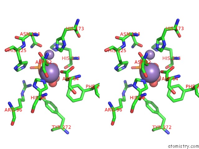
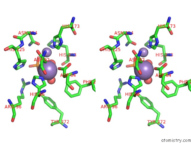
Manganese binding site 3 out of 4 in 4mov

Go back to Manganese Binding Sites List in 4mov
Manganese binding site 3 out of 4 in the 1.45 A Resolution Crystal Structure of Protein Phosphatase 1


Mono view


Stereo pair view

A full contact list of Manganese with other atoms in the Mn binding site number 3 of 1.45 A Resolution Crystal Structure of Protein Phosphatase 1 within 5.0Å range:
probe atom residue distance (Å) B Occ
B:Mn401

b:9.8
occ:1.00
OD1 B:ASN124 2.1 10.4 1.0
O1 B:PO4403 2.2 11.7 0.8
OD2 B:ASP92 2.2 9.3 1.0
NE2 B:HIS173 2.2 9.6 1.0
ND1 B:HIS248 2.2 10.6 1.0
O B:HOH508 2.3 13.6 1.0
CE1 B:HIS248 3.1 11.5 1.0
CG B:ASP92 3.2 9.5 1.0
CG B:ASN124 3.2 10.0 1.0
CE1 B:HIS173 3.2 10.2 1.0
MN B:MN402 3.2 13.6 0.5
CD2 B:HIS173 3.2 9.8 1.0
CG B:HIS248 3.4 10.6 1.0
P B:PO4403 3.4 18.5 0.8
OD1 B:ASP92 3.5 9.4 1.0
ND2 B:ASN124 3.6 11.3 1.0
O3 B:PO4403 3.7 20.3 0.8
CA B:HIS248 3.8 11.0 1.0
CB B:HIS248 3.9 10.0 1.0
OD2 B:ASP64 3.9 13.2 1.0
O2 B:PO4403 4.1 21.7 0.8
O B:HIS248 4.1 20.6 1.0
CD2 B:HIS125 4.2 9.8 1.0
NE2 B:HIS248 4.2 12.6 1.0
ND1 B:HIS173 4.3 8.4 1.0
CG B:HIS173 4.4 8.0 1.0
CB B:ASP92 4.4 8.1 1.0
CD2 B:HIS248 4.4 13.3 1.0
C B:HIS248 4.5 13.6 1.0
CB B:ASN124 4.5 9.7 1.0
N B:ASN124 4.5 8.7 1.0
O4 B:PO4403 4.6 20.1 0.8
O B:LEU205 4.7 12.1 1.0
NE2 B:HIS125 4.8 10.9 1.0
N B:HIS248 4.9 10.1 1.0
CG B:ASP64 4.9 9.3 1.0
NE2 B:HIS66 4.9 10.6 1.0
CA B:ASN124 5.0 9.3 1.0

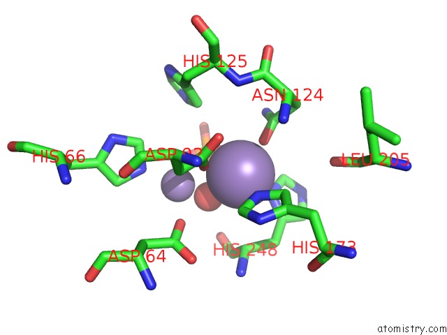
Manganese binding site 4 out of 4 in 4mov

Go back to Manganese Binding Sites List in 4mov
Manganese binding site 4 out of 4 in the 1.45 A Resolution Crystal Structure of Protein Phosphatase 1


Mono view


Stereo pair view

A full contact list of Manganese with other atoms in the Mn binding site number 4 of 1.45 A Resolution Crystal Structure of Protein Phosphatase 1 within 5.0Å range:
probe atom residue distance (Å) B Occ
B:Mn402

b:13.6
occ:0.50
O B:HOH508 1.8 13.6 1.0
NE2 B:HIS66 2.1 10.6 1.0
O3 B:PO4403 2.1 20.3 0.8
O B:HOH685 2.2 28.5 1.0
OD2 B:ASP92 2.3 9.3 1.0
OD2 B:ASP64 2.3 13.2 1.0
CE1 B:HIS66 3.0 9.0 1.0
CD2 B:HIS66 3.1 9.8 1.0
MN B:MN401 3.2 9.8 1.0
P B:PO4403 3.3 18.5 0.8
CG B:ASP92 3.4 9.5 1.0
O1 B:PO4403 3.4 11.7 0.8
CG B:ASP64 3.5 9.3 1.0
CB B:ASP92 3.7 8.1 1.0
O2 B:PO4403 3.9 21.7 0.8
O B:HIS248 4.0 20.6 1.0
CB B:ASP64 4.2 9.0 1.0
ND1 B:HIS66 4.2 8.5 1.0
NH1 B:ARG96 4.2 26.3 1.0
CG B:HIS66 4.2 7.8 1.0
CD2 B:HIS125 4.3 9.8 1.0
CE2 B:PHE267 4.3 10.6 1.0
OH B:TYR272 4.4 34.8 1.0
OD1 B:ASP92 4.5 9.4 1.0
OD1 B:ASP64 4.5 10.9 1.0
O4 B:PO4403 4.5 20.1 0.8
NE2 B:HIS125 4.5 10.9 1.0
NE2 B:HIS173 4.5 9.6 1.0
CE1 B:HIS173 4.6 10.2 1.0
CA B:HIS248 4.7 11.0 1.0
C B:HIS248 4.7 13.6 1.0
ND1 B:HIS248 4.8 10.6 1.0
CZ B:PHE267 4.9 12.5 1.0
OD1 B:ASN124 4.9 10.4 1.0

Reference:

M.S.Choy, M.Hieke, G.S.Kumar, G.R.Lewis, K.R.Gonzalez-Dewhitt, R.P.Kessler, B.J.Stein, M.Hessenberger, A.C.Nairn, W.Peti, R.Page. Understanding the Antagonism of Retinoblastoma Protein Dephosphorylation By Pnuts Provides Insights Into the PP1 Regulatory Code. Proc.Natl.Acad.Sci.Usa V. 111 4097 2014.
ISSN: ISSN 0027-8424
PubMed: 24591642
DOI: 10.1073/PNAS.1317395111
Page generated: Sat Oct 5 20:23:35 2024

Last articles

K in 6DP2
K in 6DP1
K in 6DP0
K in 6DOR
K in 6DOS
K in 6DOZ
K in 6DOQ
K in 6DOO
K in 6DON
K in 6DOM
© Copyright 2008-2020 by atomistry.com
Home   |    Site Map   |    Copyright   |    Contact us   |    Privacy