Manganese »
PDB 4lta-4mu3 »
4m1i »
Manganese in PDB 4m1i: X-Ray Crystal Structure of Chlamydia Trachomatis Mn(II)Fe(II)-Nrdb
Enzymatic activity of X-Ray Crystal Structure of Chlamydia Trachomatis Mn(II)Fe(II)-Nrdb
All present enzymatic activity of X-Ray Crystal Structure of Chlamydia Trachomatis Mn(II)Fe(II)-Nrdb:
1.17.4.1;
1.17.4.1;
Protein crystallography data
The structure of X-Ray Crystal Structure of Chlamydia Trachomatis Mn(II)Fe(II)-Nrdb, PDB code: 4m1i
was solved by
A.K.Boal,
A.C.Rosenzweig,
with X-Ray Crystallography technique. A brief refinement statistics is given in the table below:
| Resolution Low / High (Å) | 30.85 / 1.80 |
| Space group | P 1 21 1 |
| Cell size a, b, c (Å), α, β, γ (°) | 74.897, 97.455, 99.315, 90.00, 97.77, 90.00 |
| R / Rfree (%) | 17.2 / 20.2 |
Other elements in 4m1i:
The structure of X-Ray Crystal Structure of Chlamydia Trachomatis Mn(II)Fe(II)-Nrdb also contains other interesting chemical elements:
| Iron | (Fe) | 4 atoms |
Manganese Binding Sites:
The binding sites of Manganese atom in the X-Ray Crystal Structure of Chlamydia Trachomatis Mn(II)Fe(II)-Nrdb
(pdb code 4m1i). This binding sites where shown within
5.0 Angstroms radius around Manganese atom.
In total 4 binding sites of Manganese where determined in the X-Ray Crystal Structure of Chlamydia Trachomatis Mn(II)Fe(II)-Nrdb, PDB code: 4m1i:
Jump to Manganese binding site number: 1; 2; 3; 4;
In total 4 binding sites of Manganese where determined in the X-Ray Crystal Structure of Chlamydia Trachomatis Mn(II)Fe(II)-Nrdb, PDB code: 4m1i:
Jump to Manganese binding site number: 1; 2; 3; 4;
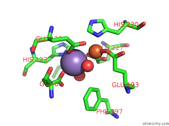
Manganese binding site 1 out of 4 in 4m1i
Go back to
Manganese binding site 1 out
of 4 in the X-Ray Crystal Structure of Chlamydia Trachomatis Mn(II)Fe(II)-Nrdb
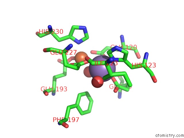
Mono view
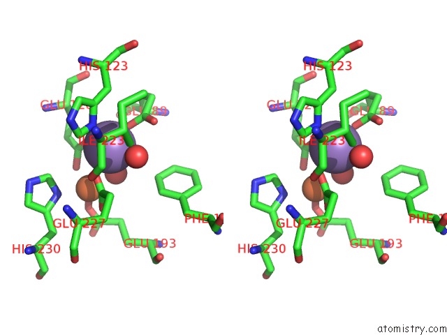
Stereo pair view
Mono view
Stereo pair view
A full contact list of Manganese with other atoms in the Mn binding
site number 1 of X-Ray Crystal Structure of Chlamydia Trachomatis Mn(II)Fe(II)-Nrdb within 5.0Å range:
|
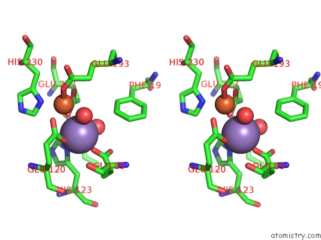
Manganese binding site 2 out of 4 in 4m1i
Go back to
Manganese binding site 2 out
of 4 in the X-Ray Crystal Structure of Chlamydia Trachomatis Mn(II)Fe(II)-Nrdb
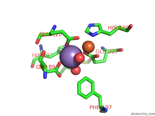
Mono view
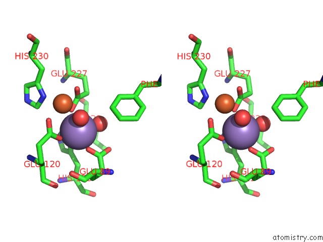
Stereo pair view
Mono view
Stereo pair view
A full contact list of Manganese with other atoms in the Mn binding
site number 2 of X-Ray Crystal Structure of Chlamydia Trachomatis Mn(II)Fe(II)-Nrdb within 5.0Å range:
|
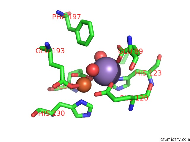
Manganese binding site 3 out of 4 in 4m1i
Go back to
Manganese binding site 3 out
of 4 in the X-Ray Crystal Structure of Chlamydia Trachomatis Mn(II)Fe(II)-Nrdb
Mono view
Stereo pair view
Mono view
Stereo pair view
A full contact list of Manganese with other atoms in the Mn binding
site number 3 of X-Ray Crystal Structure of Chlamydia Trachomatis Mn(II)Fe(II)-Nrdb within 5.0Å range:
|
Manganese binding site 4 out of 4 in 4m1i
Go back to
Manganese binding site 4 out
of 4 in the X-Ray Crystal Structure of Chlamydia Trachomatis Mn(II)Fe(II)-Nrdb
Mono view
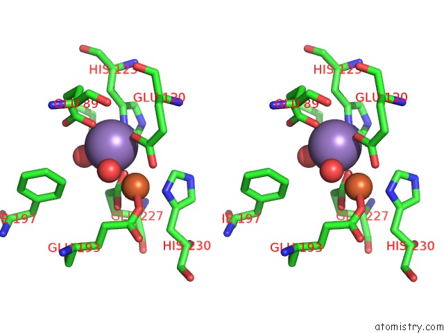
Stereo pair view
Mono view
Stereo pair view
A full contact list of Manganese with other atoms in the Mn binding
site number 4 of X-Ray Crystal Structure of Chlamydia Trachomatis Mn(II)Fe(II)-Nrdb within 5.0Å range:
|
Reference:
L.M.Dassama,
C.Krebs,
J.M.Bollinger,
A.C.Rosenzweig,
A.K.Boal.
Structural Basis For Assembly of the Mn(IV)/Fe(III) Cofactor in the Class Ic Ribonucleotide Reductase From Chlamydia Trachomatis. Biochemistry V. 52 6424 2013.
ISSN: ISSN 0006-2960
PubMed: 23924396
DOI: 10.1021/BI400819X
Page generated: Sat Oct 5 20:18:00 2024
ISSN: ISSN 0006-2960
PubMed: 23924396
DOI: 10.1021/BI400819X
Last articles
Mg in 3SRFMg in 3SRD
Mg in 3SR0
Mg in 3SPY
Mg in 3SQX
Mg in 3SQS
Mg in 3SQW
Mg in 3SPT
Mg in 3SPR
Mg in 3SP5