Manganese »
PDB 4gwc-4ilk »
4icm »
Manganese in PDB 4icm: Crystal Structure of 5-Carboxyvanillate Decarboxylase Ligw From Sphingomonas Paucimobilis
Protein crystallography data
The structure of Crystal Structure of 5-Carboxyvanillate Decarboxylase Ligw From Sphingomonas Paucimobilis, PDB code: 4icm
was solved by
A.A.Fedorov,
E.V.Fedorov,
A.Vladimirova,
F.M.Raushel,
S.C.Almo,
with X-Ray Crystallography technique. A brief refinement statistics is given in the table below:
| Resolution Low / High (Å) | 38.51 / 1.82 |
| Space group | P 1 |
| Cell size a, b, c (Å), α, β, γ (°) | 80.817, 96.897, 97.004, 109.58, 90.48, 111.66 |
| R / Rfree (%) | 16.3 / 20.5 |
Manganese Binding Sites:
The binding sites of Manganese atom in the Crystal Structure of 5-Carboxyvanillate Decarboxylase Ligw From Sphingomonas Paucimobilis
(pdb code 4icm). This binding sites where shown within
5.0 Angstroms radius around Manganese atom.
In total 8 binding sites of Manganese where determined in the Crystal Structure of 5-Carboxyvanillate Decarboxylase Ligw From Sphingomonas Paucimobilis, PDB code: 4icm:
Jump to Manganese binding site number: 1; 2; 3; 4; 5; 6; 7; 8;
In total 8 binding sites of Manganese where determined in the Crystal Structure of 5-Carboxyvanillate Decarboxylase Ligw From Sphingomonas Paucimobilis, PDB code: 4icm:
Jump to Manganese binding site number: 1; 2; 3; 4; 5; 6; 7; 8;
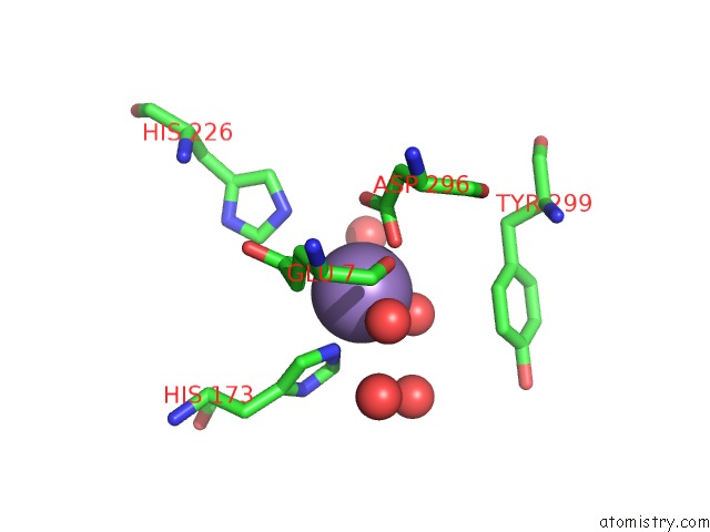
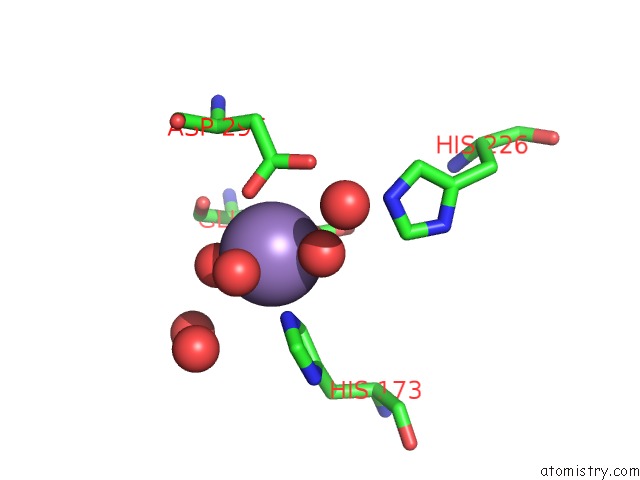
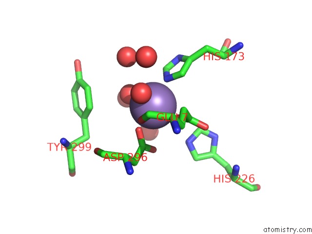
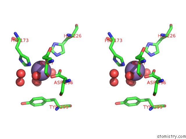
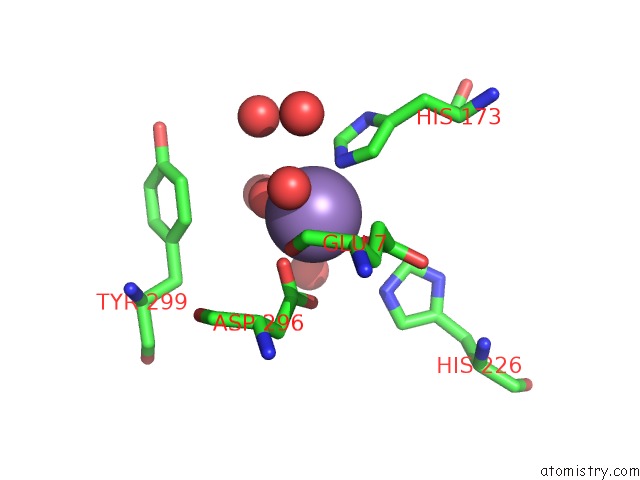
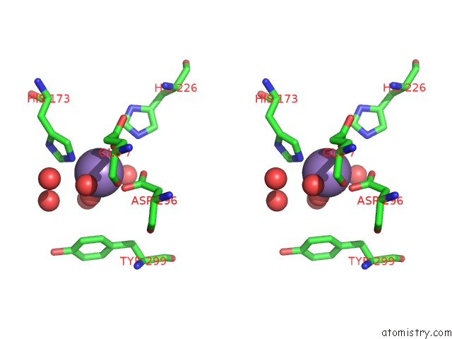
Manganese binding site 1 out of 8 in 4icm
Go back to
Manganese binding site 1 out
of 8 in the Crystal Structure of 5-Carboxyvanillate Decarboxylase Ligw From Sphingomonas Paucimobilis
Mono view
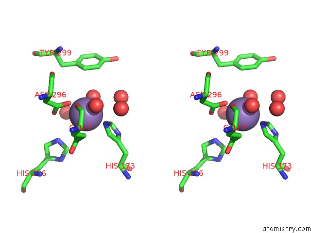
Stereo pair view
Mono view
Stereo pair view
A full contact list of Manganese with other atoms in the Mn binding
site number 1 of Crystal Structure of 5-Carboxyvanillate Decarboxylase Ligw From Sphingomonas Paucimobilis within 5.0Å range:
|
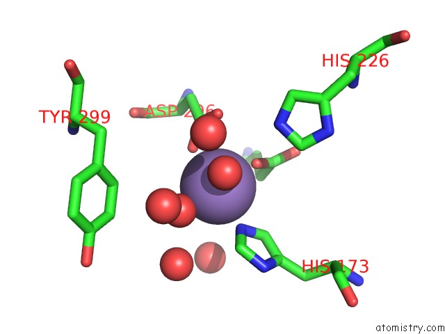
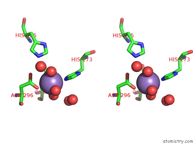
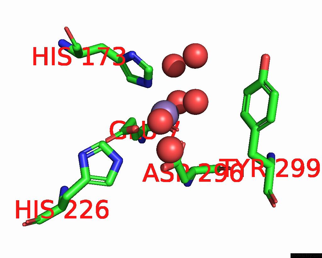
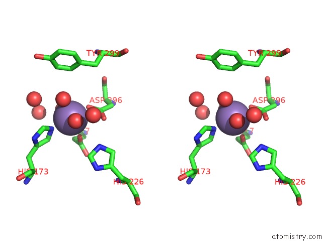
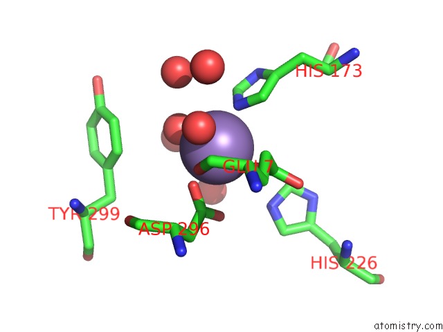
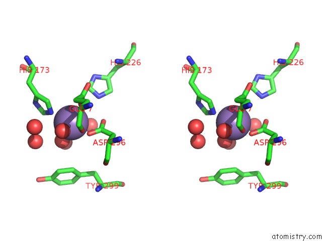
Manganese binding site 2 out of 8 in 4icm
Go back to
Manganese binding site 2 out
of 8 in the Crystal Structure of 5-Carboxyvanillate Decarboxylase Ligw From Sphingomonas Paucimobilis
Mono view
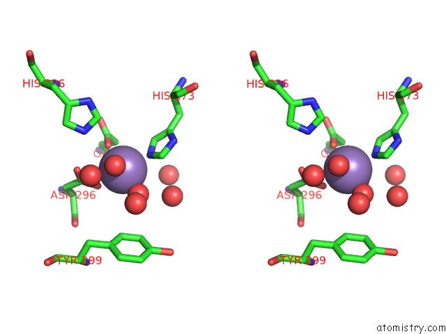
Stereo pair view
Mono view
Stereo pair view
A full contact list of Manganese with other atoms in the Mn binding
site number 2 of Crystal Structure of 5-Carboxyvanillate Decarboxylase Ligw From Sphingomonas Paucimobilis within 5.0Å range:
|
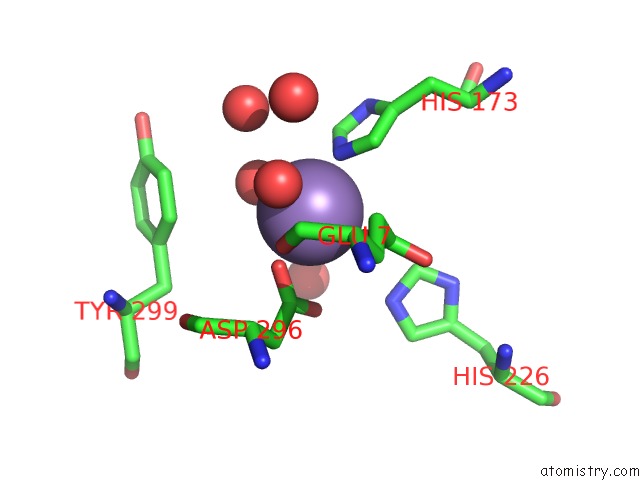
Manganese binding site 3 out of 8 in 4icm
Go back to
Manganese binding site 3 out
of 8 in the Crystal Structure of 5-Carboxyvanillate Decarboxylase Ligw From Sphingomonas Paucimobilis
Mono view
Stereo pair view
Mono view
Stereo pair view
A full contact list of Manganese with other atoms in the Mn binding
site number 3 of Crystal Structure of 5-Carboxyvanillate Decarboxylase Ligw From Sphingomonas Paucimobilis within 5.0Å range:
|
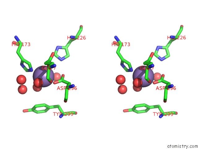
Manganese binding site 4 out of 8 in 4icm
Go back to
Manganese binding site 4 out
of 8 in the Crystal Structure of 5-Carboxyvanillate Decarboxylase Ligw From Sphingomonas Paucimobilis
Mono view
Stereo pair view
Mono view
Stereo pair view
A full contact list of Manganese with other atoms in the Mn binding
site number 4 of Crystal Structure of 5-Carboxyvanillate Decarboxylase Ligw From Sphingomonas Paucimobilis within 5.0Å range:
|
Manganese binding site 5 out of 8 in 4icm
Go back to
Manganese binding site 5 out
of 8 in the Crystal Structure of 5-Carboxyvanillate Decarboxylase Ligw From Sphingomonas Paucimobilis
Mono view
Stereo pair view
Mono view
Stereo pair view
A full contact list of Manganese with other atoms in the Mn binding
site number 5 of Crystal Structure of 5-Carboxyvanillate Decarboxylase Ligw From Sphingomonas Paucimobilis within 5.0Å range:
|
Manganese binding site 6 out of 8 in 4icm
Go back to
Manganese binding site 6 out
of 8 in the Crystal Structure of 5-Carboxyvanillate Decarboxylase Ligw From Sphingomonas Paucimobilis
Mono view
Stereo pair view
Mono view
Stereo pair view
A full contact list of Manganese with other atoms in the Mn binding
site number 6 of Crystal Structure of 5-Carboxyvanillate Decarboxylase Ligw From Sphingomonas Paucimobilis within 5.0Å range:
|
Manganese binding site 7 out of 8 in 4icm
Go back to
Manganese binding site 7 out
of 8 in the Crystal Structure of 5-Carboxyvanillate Decarboxylase Ligw From Sphingomonas Paucimobilis
Mono view
Stereo pair view
Mono view
Stereo pair view
A full contact list of Manganese with other atoms in the Mn binding
site number 7 of Crystal Structure of 5-Carboxyvanillate Decarboxylase Ligw From Sphingomonas Paucimobilis within 5.0Å range:
|
Manganese binding site 8 out of 8 in 4icm
Go back to
Manganese binding site 8 out
of 8 in the Crystal Structure of 5-Carboxyvanillate Decarboxylase Ligw From Sphingomonas Paucimobilis
Mono view
Stereo pair view
Mono view
Stereo pair view
A full contact list of Manganese with other atoms in the Mn binding
site number 8 of Crystal Structure of 5-Carboxyvanillate Decarboxylase Ligw From Sphingomonas Paucimobilis within 5.0Å range:
|
Reference:
A.A.Fedorov,
E.V.Fedorov,
A.Vladimirova,
F.M.Raushel,
S.C.Almo.
Crystal Structure of 5-Carboxyvanillate Decarboxylase Ligw From Sphingomonas Paucimobilis To Be Published.
Page generated: Sat Oct 5 19:47:28 2024
Last articles
Ca in 3OJYCa in 3OM2
Ca in 3OLI
Ca in 3OLG
Ca in 3OLE
Ca in 3OLD
Ca in 3OJT
Ca in 3OJC
Ca in 3OIW
Ca in 3OJN