Manganese »
PDB 4gwc-4ilk »
4hx4 »
Manganese in PDB 4hx4: Structure of Mntr Mutant E11K Complexed with MN2+
Protein crystallography data
The structure of Structure of Mntr Mutant E11K Complexed with MN2+, PDB code: 4hx4
was solved by
A.Glasfeld,
A.Mcguire,
with X-Ray Crystallography technique. A brief refinement statistics is given in the table below:
| Resolution Low / High (Å) | 30.33 / 1.65 |
| Space group | P 1 21 1 |
| Cell size a, b, c (Å), α, β, γ (°) | 49.427, 46.081, 74.892, 90.00, 92.96, 90.00 |
| R / Rfree (%) | 23.4 / 26.4 |
Manganese Binding Sites:
The binding sites of Manganese atom in the Structure of Mntr Mutant E11K Complexed with MN2+
(pdb code 4hx4). This binding sites where shown within
5.0 Angstroms radius around Manganese atom.
In total 2 binding sites of Manganese where determined in the Structure of Mntr Mutant E11K Complexed with MN2+, PDB code: 4hx4:
Jump to Manganese binding site number: 1; 2;
In total 2 binding sites of Manganese where determined in the Structure of Mntr Mutant E11K Complexed with MN2+, PDB code: 4hx4:
Jump to Manganese binding site number: 1; 2;
Manganese binding site 1 out of 2 in 4hx4
Go back to
Manganese binding site 1 out
of 2 in the Structure of Mntr Mutant E11K Complexed with MN2+
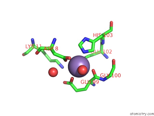
Mono view
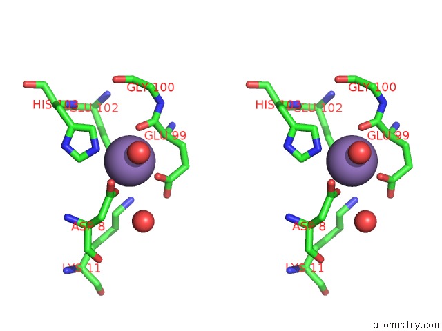
Stereo pair view
Mono view
Stereo pair view
A full contact list of Manganese with other atoms in the Mn binding
site number 1 of Structure of Mntr Mutant E11K Complexed with MN2+ within 5.0Å range:
|
Manganese binding site 2 out of 2 in 4hx4
Go back to
Manganese binding site 2 out
of 2 in the Structure of Mntr Mutant E11K Complexed with MN2+
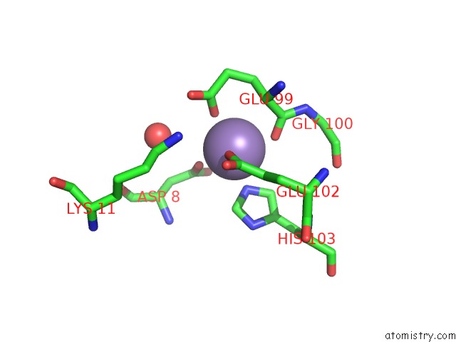
Mono view
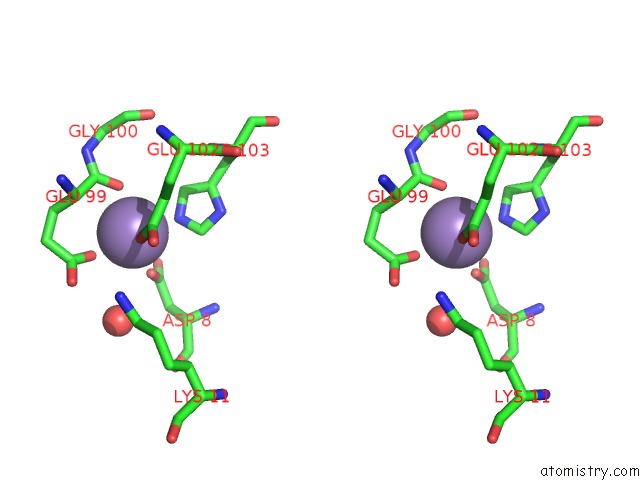
Stereo pair view
Mono view
Stereo pair view
A full contact list of Manganese with other atoms in the Mn binding
site number 2 of Structure of Mntr Mutant E11K Complexed with MN2+ within 5.0Å range:
|
Reference:
A.M.Mcguire,
B.J.Cuthbert,
Z.Ma,
K.D.Grauer-Gray,
M.Brunjes Brophy,
K.A.Spear,
S.Soonsanga,
J.I.Kliegman,
S.L.Griner,
J.D.Helmann,
A.Glasfeld.
Roles of the A and C Sites in the Manganese-Specific Activation of Mntr. Biochemistry V. 52 701 2013.
ISSN: ISSN 0006-2960
PubMed: 23298157
DOI: 10.1021/BI301550T
Page generated: Sat Oct 5 19:44:50 2024
ISSN: ISSN 0006-2960
PubMed: 23298157
DOI: 10.1021/BI301550T
Last articles
Ca in 3MVSCa in 3MX9
Ca in 3MW3
Ca in 3MW4
Ca in 3MVV
Ca in 3MSF
Ca in 3MSN
Ca in 3MT5
Ca in 3MSE
Ca in 3MSA