Manganese »
PDB 4fo7-4gv9 »
4gik »
Manganese in PDB 4gik: Crystal Structure of Pseudouridine Monophosphate Glycosidase/Linear R5P Adduct
Protein crystallography data
The structure of Crystal Structure of Pseudouridine Monophosphate Glycosidase/Linear R5P Adduct, PDB code: 4gik
was solved by
S.Huang,
N.Mahanta,
T.P.Begley,
S.E.Ealick,
with X-Ray Crystallography technique. A brief refinement statistics is given in the table below:
| Resolution Low / High (Å) | 41.20 / 2.19 |
| Space group | P 21 21 21 |
| Cell size a, b, c (Å), α, β, γ (°) | 61.347, 116.496, 132.153, 90.00, 90.00, 90.00 |
| R / Rfree (%) | 17.9 / 23.1 |
Manganese Binding Sites:
The binding sites of Manganese atom in the Crystal Structure of Pseudouridine Monophosphate Glycosidase/Linear R5P Adduct
(pdb code 4gik). This binding sites where shown within
5.0 Angstroms radius around Manganese atom.
In total 3 binding sites of Manganese where determined in the Crystal Structure of Pseudouridine Monophosphate Glycosidase/Linear R5P Adduct, PDB code: 4gik:
Jump to Manganese binding site number: 1; 2; 3;
In total 3 binding sites of Manganese where determined in the Crystal Structure of Pseudouridine Monophosphate Glycosidase/Linear R5P Adduct, PDB code: 4gik:
Jump to Manganese binding site number: 1; 2; 3;
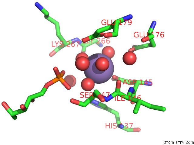
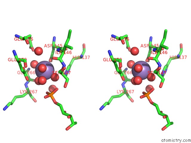
Manganese binding site 1 out of 3 in 4gik
Go back to
Manganese binding site 1 out
of 3 in the Crystal Structure of Pseudouridine Monophosphate Glycosidase/Linear R5P Adduct
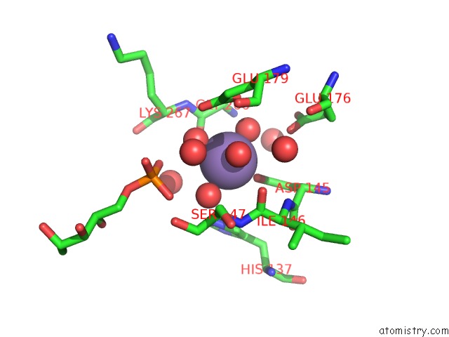
Mono view
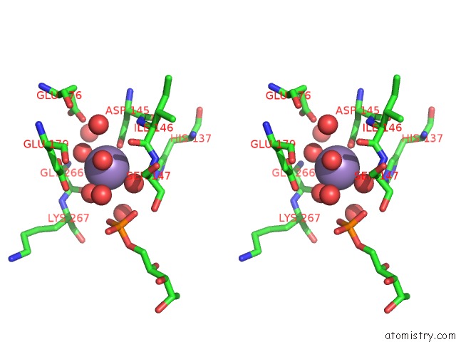
Stereo pair view
Mono view
Stereo pair view
A full contact list of Manganese with other atoms in the Mn binding
site number 1 of Crystal Structure of Pseudouridine Monophosphate Glycosidase/Linear R5P Adduct within 5.0Å range:
|
Manganese binding site 2 out of 3 in 4gik
Go back to
Manganese binding site 2 out
of 3 in the Crystal Structure of Pseudouridine Monophosphate Glycosidase/Linear R5P Adduct
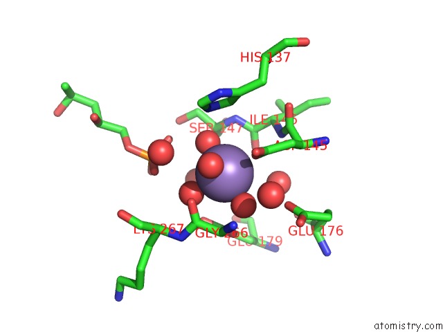
Mono view
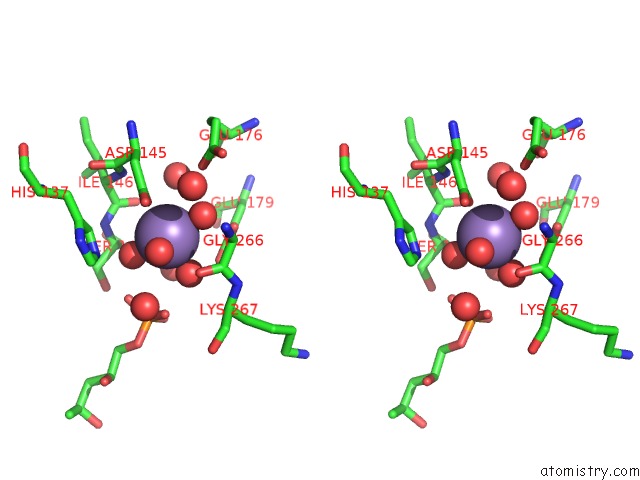
Stereo pair view
Mono view
Stereo pair view
A full contact list of Manganese with other atoms in the Mn binding
site number 2 of Crystal Structure of Pseudouridine Monophosphate Glycosidase/Linear R5P Adduct within 5.0Å range:
|
Manganese binding site 3 out of 3 in 4gik
Go back to
Manganese binding site 3 out
of 3 in the Crystal Structure of Pseudouridine Monophosphate Glycosidase/Linear R5P Adduct
Mono view
Stereo pair view
Mono view
Stereo pair view
A full contact list of Manganese with other atoms in the Mn binding
site number 3 of Crystal Structure of Pseudouridine Monophosphate Glycosidase/Linear R5P Adduct within 5.0Å range:
|
Reference:
S.Huang,
N.Mahanta,
T.P.Begley,
S.E.Ealick.
Pseudouridine Monophosphate Glycosidase: A New Glycosidase Mechanism. Biochemistry V. 51 9245 2012.
ISSN: ISSN 0006-2960
PubMed: 23066817
DOI: 10.1021/BI3006829
Page generated: Sat Oct 5 19:31:03 2024
ISSN: ISSN 0006-2960
PubMed: 23066817
DOI: 10.1021/BI3006829
Last articles
Mg in 3CK4Mg in 3CJO
Mg in 3CJ8
Mg in 3CIK
Mg in 3CIN
Mg in 3CIP
Mg in 3CIF
Mg in 3CFX
Mg in 3CI5
Mg in 3CI4