Manganese »
PDB 4fo7-4gv9 »
4ggf »
Manganese in PDB 4ggf: Crystal Structure of MN2+ Bound Calprotectin
Protein crystallography data
The structure of Crystal Structure of MN2+ Bound Calprotectin, PDB code: 4ggf
was solved by
S.M.Damo,
G.Fritz,
with X-Ray Crystallography technique. A brief refinement statistics is given in the table below:
| Resolution Low / High (Å) | 47.64 / 1.60 |
| Space group | P 21 21 2 |
| Cell size a, b, c (Å), α, β, γ (°) | 82.027, 217.002, 53.022, 90.00, 90.00, 90.00 |
| R / Rfree (%) | 17.7 / 20.2 |
Other elements in 4ggf:
The structure of Crystal Structure of MN2+ Bound Calprotectin also contains other interesting chemical elements:
| Calcium | (Ca) | 12 atoms |
Manganese Binding Sites:
The binding sites of Manganese atom in the Crystal Structure of MN2+ Bound Calprotectin
(pdb code 4ggf). This binding sites where shown within
5.0 Angstroms radius around Manganese atom.
In total 7 binding sites of Manganese where determined in the Crystal Structure of MN2+ Bound Calprotectin, PDB code: 4ggf:
Jump to Manganese binding site number: 1; 2; 3; 4; 5; 6; 7;
In total 7 binding sites of Manganese where determined in the Crystal Structure of MN2+ Bound Calprotectin, PDB code: 4ggf:
Jump to Manganese binding site number: 1; 2; 3; 4; 5; 6; 7;
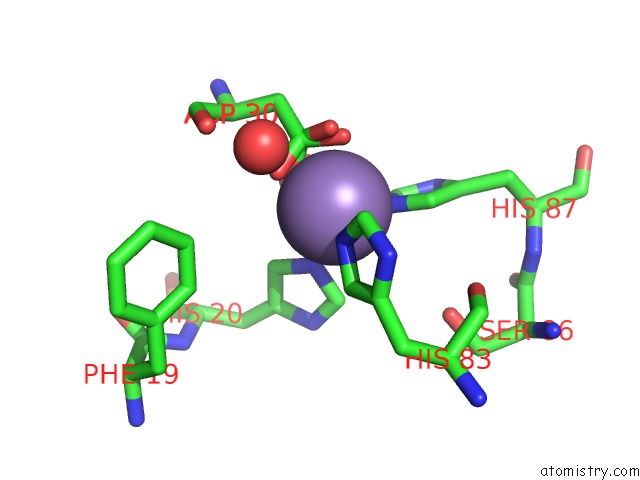
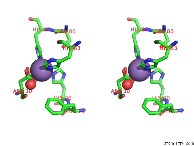
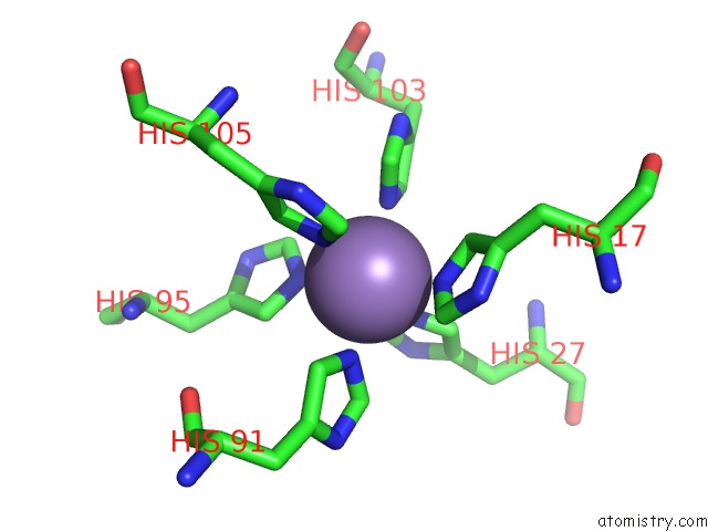
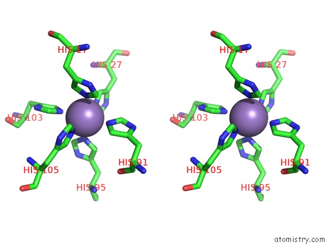
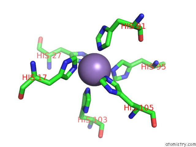
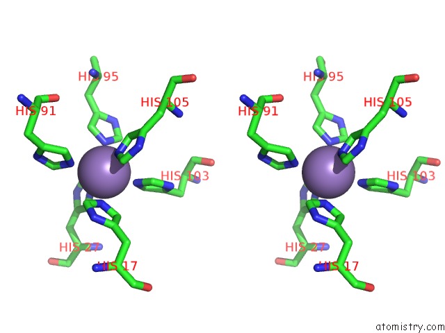
Manganese binding site 1 out of 7 in 4ggf
Go back to
Manganese binding site 1 out
of 7 in the Crystal Structure of MN2+ Bound Calprotectin
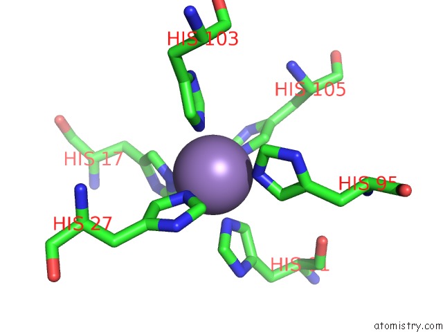
Mono view
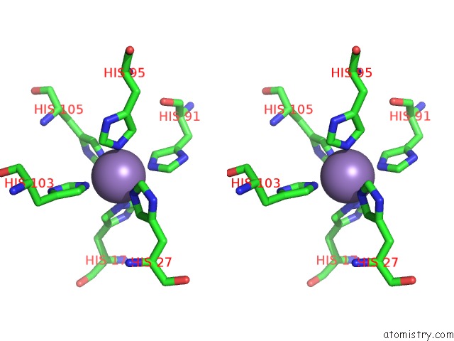
Stereo pair view
Mono view
Stereo pair view
A full contact list of Manganese with other atoms in the Mn binding
site number 1 of Crystal Structure of MN2+ Bound Calprotectin within 5.0Å range:
|
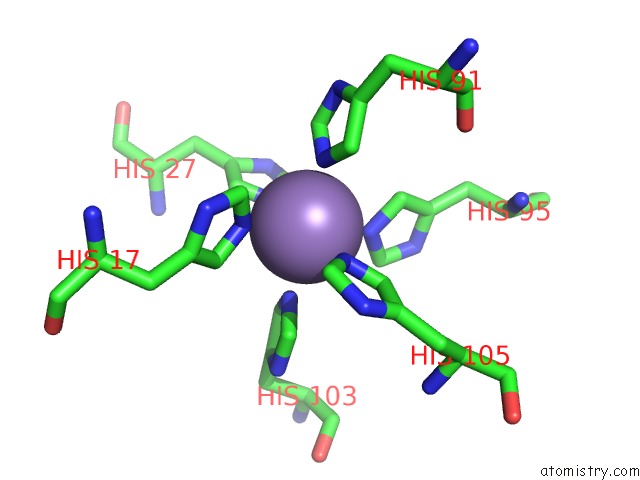
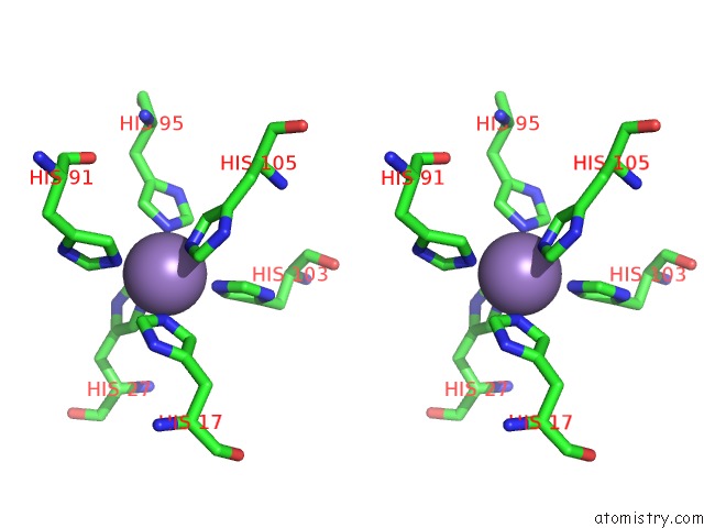
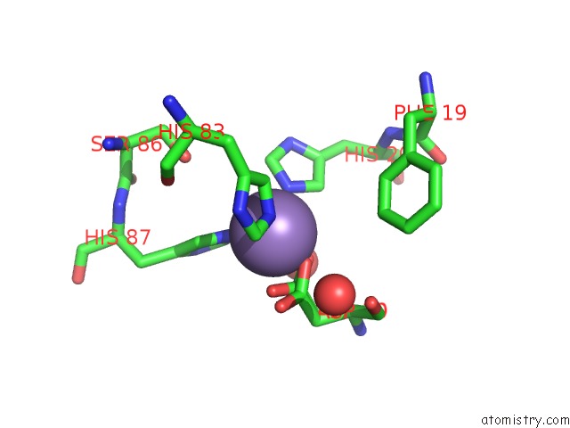
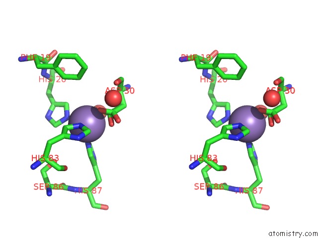
Manganese binding site 2 out of 7 in 4ggf
Go back to
Manganese binding site 2 out
of 7 in the Crystal Structure of MN2+ Bound Calprotectin
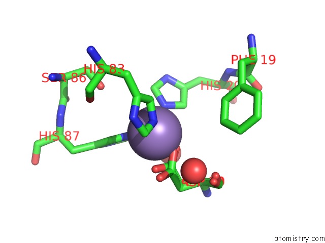
Mono view
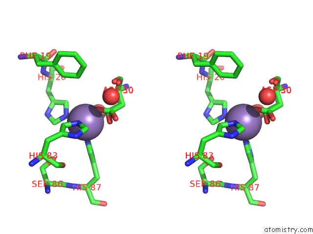
Stereo pair view
Mono view
Stereo pair view
A full contact list of Manganese with other atoms in the Mn binding
site number 2 of Crystal Structure of MN2+ Bound Calprotectin within 5.0Å range:
|
Manganese binding site 3 out of 7 in 4ggf
Go back to
Manganese binding site 3 out
of 7 in the Crystal Structure of MN2+ Bound Calprotectin
Mono view
Stereo pair view
Mono view
Stereo pair view
A full contact list of Manganese with other atoms in the Mn binding
site number 3 of Crystal Structure of MN2+ Bound Calprotectin within 5.0Å range:
|
Manganese binding site 4 out of 7 in 4ggf
Go back to
Manganese binding site 4 out
of 7 in the Crystal Structure of MN2+ Bound Calprotectin
Mono view
Stereo pair view
Mono view
Stereo pair view
A full contact list of Manganese with other atoms in the Mn binding
site number 4 of Crystal Structure of MN2+ Bound Calprotectin within 5.0Å range:
|
Manganese binding site 5 out of 7 in 4ggf
Go back to
Manganese binding site 5 out
of 7 in the Crystal Structure of MN2+ Bound Calprotectin
Mono view
Stereo pair view
Mono view
Stereo pair view
A full contact list of Manganese with other atoms in the Mn binding
site number 5 of Crystal Structure of MN2+ Bound Calprotectin within 5.0Å range:
|
Manganese binding site 6 out of 7 in 4ggf
Go back to
Manganese binding site 6 out
of 7 in the Crystal Structure of MN2+ Bound Calprotectin
Mono view
Stereo pair view
Mono view
Stereo pair view
A full contact list of Manganese with other atoms in the Mn binding
site number 6 of Crystal Structure of MN2+ Bound Calprotectin within 5.0Å range:
|
Manganese binding site 7 out of 7 in 4ggf
Go back to
Manganese binding site 7 out
of 7 in the Crystal Structure of MN2+ Bound Calprotectin
Mono view
Stereo pair view
Mono view
Stereo pair view
A full contact list of Manganese with other atoms in the Mn binding
site number 7 of Crystal Structure of MN2+ Bound Calprotectin within 5.0Å range:
|
Reference:
S.M.Damo,
T.E.Kehl-Fie,
N.Sugitani,
M.E.Holt,
S.Rathi,
W.J.Murphy,
Y.Zhang,
C.Betz,
L.Hench,
G.Fritz,
E.P.Skaar,
W.J.Chazin.
Molecular Basis For Manganese Sequestration By Calprotectin and Roles in the Innate Immune Response to Invading Bacterial Pathogens. Proc.Natl.Acad.Sci.Usa V. 110 3841 2013.
ISSN: ISSN 0027-8424
PubMed: 23431180
DOI: 10.1073/PNAS.1220341110
Page generated: Sat Oct 5 19:30:09 2024
ISSN: ISSN 0027-8424
PubMed: 23431180
DOI: 10.1073/PNAS.1220341110
Last articles
K in 4TM0K in 4TMZ
K in 4TLZ
K in 4TLX
K in 4RVO
K in 4RVN
K in 4TKX
K in 4RUM
K in 4RUF
K in 4RUE