Manganese »
PDB 4ee3-4fo6 »
4fix »
Manganese in PDB 4fix: Crystal Structure of GLFT2
Protein crystallography data
The structure of Crystal Structure of GLFT2, PDB code: 4fix
was solved by
R.W.Wheatley,
R.B.Zheng,
T.L.Lowary,
K.K.S.Ng,
with X-Ray Crystallography technique. A brief refinement statistics is given in the table below:
| Resolution Low / High (Å) | 40.00 / 2.45 |
| Space group | P 4 21 2 |
| Cell size a, b, c (Å), α, β, γ (°) | 150.791, 150.791, 148.016, 90.00, 90.00, 90.00 |
| R / Rfree (%) | 18.5 / 22.2 |
Manganese Binding Sites:
The binding sites of Manganese atom in the Crystal Structure of GLFT2
(pdb code 4fix). This binding sites where shown within
5.0 Angstroms radius around Manganese atom.
In total 2 binding sites of Manganese where determined in the Crystal Structure of GLFT2, PDB code: 4fix:
Jump to Manganese binding site number: 1; 2;
In total 2 binding sites of Manganese where determined in the Crystal Structure of GLFT2, PDB code: 4fix:
Jump to Manganese binding site number: 1; 2;
Manganese binding site 1 out of 2 in 4fix
Go back to
Manganese binding site 1 out
of 2 in the Crystal Structure of GLFT2
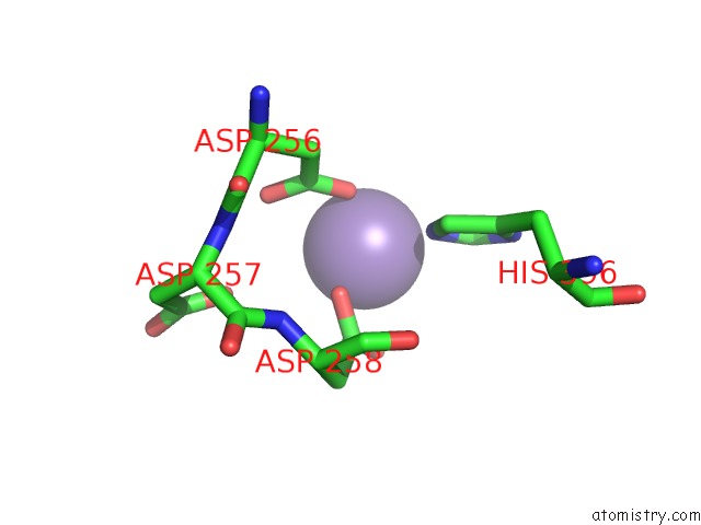
Mono view
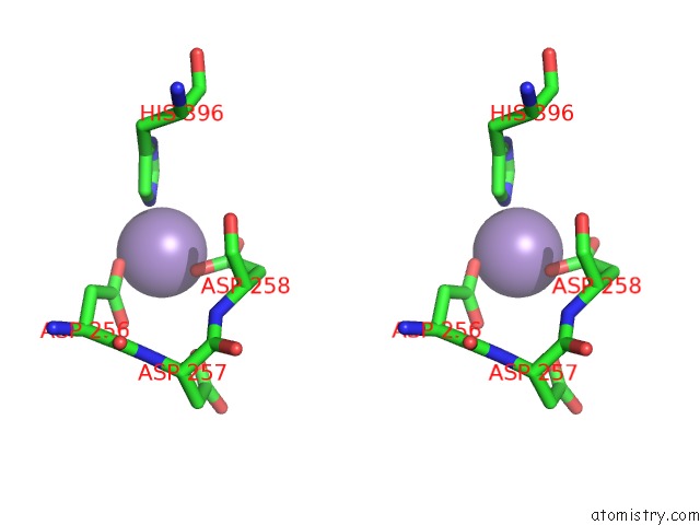
Stereo pair view
Mono view
Stereo pair view
A full contact list of Manganese with other atoms in the Mn binding
site number 1 of Crystal Structure of GLFT2 within 5.0Å range:
|
Manganese binding site 2 out of 2 in 4fix
Go back to
Manganese binding site 2 out
of 2 in the Crystal Structure of GLFT2
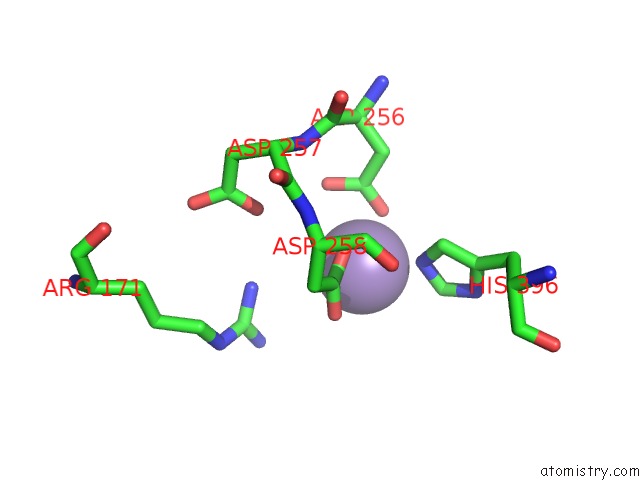
Mono view
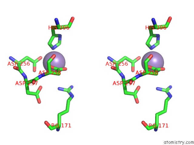
Stereo pair view
Mono view
Stereo pair view
A full contact list of Manganese with other atoms in the Mn binding
site number 2 of Crystal Structure of GLFT2 within 5.0Å range:
|
Reference:
R.W.Wheatley,
R.B.Zheng,
M.R.Richards,
T.L.Lowary,
K.K.Ng.
Tetrameric Structure of the GLFT2 Galactofuranosyltransferase Reveals A Scaffold For the Assembly of Mycobacterial Arabinogalactan. J.Biol.Chem. V. 287 28132 2012.
ISSN: ISSN 0021-9258
PubMed: 22707726
DOI: 10.1074/JBC.M112.347484
Page generated: Sat Oct 5 19:25:38 2024
ISSN: ISSN 0021-9258
PubMed: 22707726
DOI: 10.1074/JBC.M112.347484
Last articles
K in 4LF5K in 4LF4
K in 4LE2
K in 4LCU
K in 4LCA
K in 4LC4
K in 4LBX
K in 4LBG
K in 4LBE
K in 4L6A