Manganese »
PDB 4ee3-4fo6 »
4fci »
Manganese in PDB 4fci: Crystal Structure of the MN2+2-Human Arginase I-Agpa Complex
Enzymatic activity of Crystal Structure of the MN2+2-Human Arginase I-Agpa Complex
All present enzymatic activity of Crystal Structure of the MN2+2-Human Arginase I-Agpa Complex:
3.5.3.1;
3.5.3.1;
Protein crystallography data
The structure of Crystal Structure of the MN2+2-Human Arginase I-Agpa Complex, PDB code: 4fci
was solved by
E.L.D'antonio,
D.W.Christianson,
with X-Ray Crystallography technique. A brief refinement statistics is given in the table below:
| Resolution Low / High (Å) | 50.00 / 1.82 |
| Space group | P 3 |
| Cell size a, b, c (Å), α, β, γ (°) | 90.848, 90.848, 69.767, 90.00, 90.00, 120.00 |
| R / Rfree (%) | 27.9 / 31 |
Manganese Binding Sites:
The binding sites of Manganese atom in the Crystal Structure of the MN2+2-Human Arginase I-Agpa Complex
(pdb code 4fci). This binding sites where shown within
5.0 Angstroms radius around Manganese atom.
In total 4 binding sites of Manganese where determined in the Crystal Structure of the MN2+2-Human Arginase I-Agpa Complex, PDB code: 4fci:
Jump to Manganese binding site number: 1; 2; 3; 4;
In total 4 binding sites of Manganese where determined in the Crystal Structure of the MN2+2-Human Arginase I-Agpa Complex, PDB code: 4fci:
Jump to Manganese binding site number: 1; 2; 3; 4;
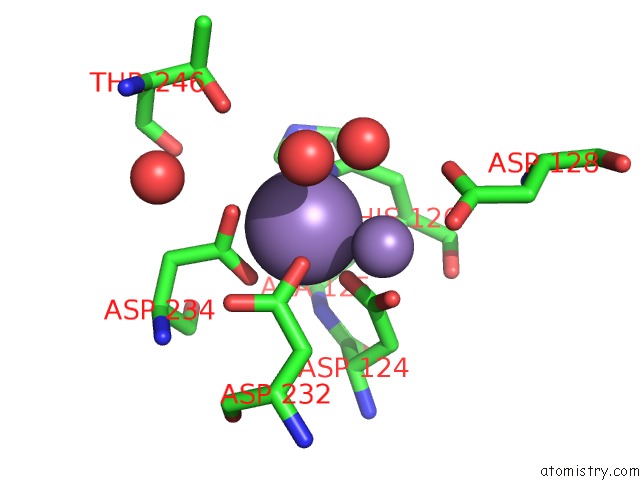
Manganese binding site 1 out of 4 in 4fci
Go back to
Manganese binding site 1 out
of 4 in the Crystal Structure of the MN2+2-Human Arginase I-Agpa Complex
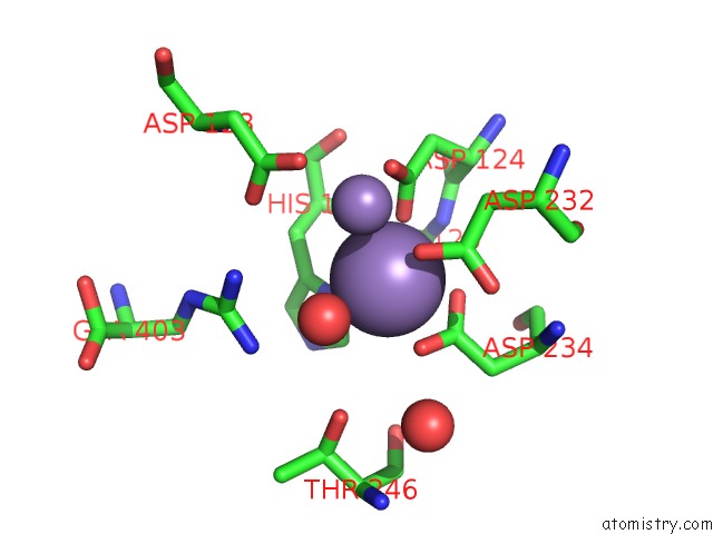
Mono view
Stereo pair view
Mono view
Stereo pair view
A full contact list of Manganese with other atoms in the Mn binding
site number 1 of Crystal Structure of the MN2+2-Human Arginase I-Agpa Complex within 5.0Å range:
|
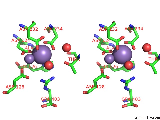
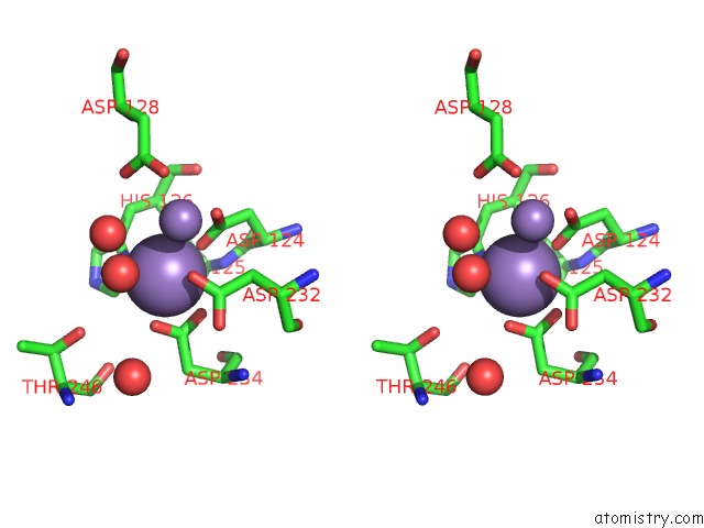
Manganese binding site 2 out of 4 in 4fci
Go back to
Manganese binding site 2 out
of 4 in the Crystal Structure of the MN2+2-Human Arginase I-Agpa Complex
Mono view
Stereo pair view
Mono view
Stereo pair view
A full contact list of Manganese with other atoms in the Mn binding
site number 2 of Crystal Structure of the MN2+2-Human Arginase I-Agpa Complex within 5.0Å range:
|
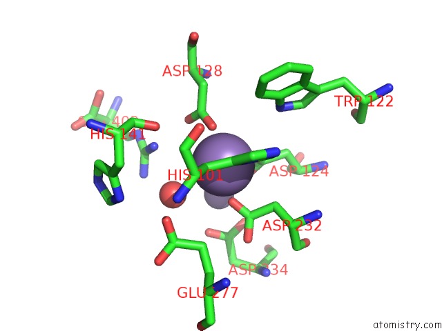
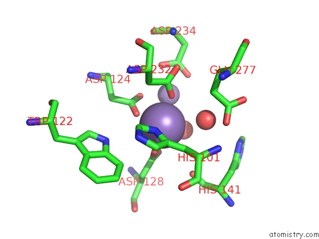
Manganese binding site 3 out of 4 in 4fci
Go back to
Manganese binding site 3 out
of 4 in the Crystal Structure of the MN2+2-Human Arginase I-Agpa Complex
Mono view
Stereo pair view
Mono view
Stereo pair view
A full contact list of Manganese with other atoms in the Mn binding
site number 3 of Crystal Structure of the MN2+2-Human Arginase I-Agpa Complex within 5.0Å range:
|
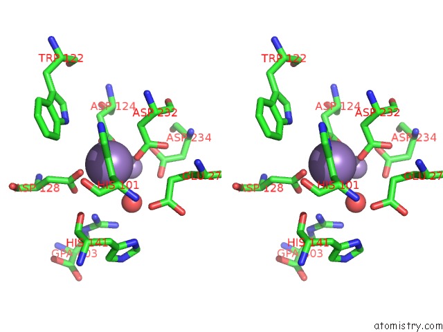
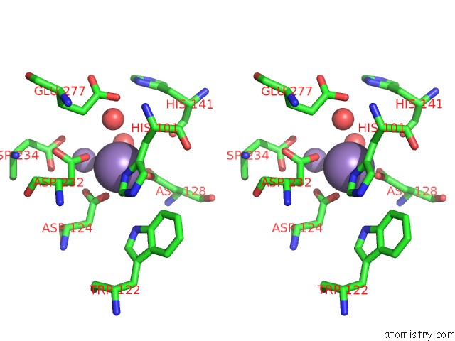
Manganese binding site 4 out of 4 in 4fci
Go back to
Manganese binding site 4 out
of 4 in the Crystal Structure of the MN2+2-Human Arginase I-Agpa Complex
Mono view
Stereo pair view
Mono view
Stereo pair view
A full contact list of Manganese with other atoms in the Mn binding
site number 4 of Crystal Structure of the MN2+2-Human Arginase I-Agpa Complex within 5.0Å range:
|
Reference:
E.L.D'antonio,
D.W.Christianson.
Binding of the Unreactive Substrate Analog L-2-Amino-3-Guanidinopropionic Acid (Dinor-L-Arginine) to Human Arginase I. Acta Crystallogr.,Sect.F V. 68 889 2012.
ISSN: ESSN 1744-3091
PubMed: 22869115
DOI: 10.1107/S1744309112027820
Page generated: Sat Oct 5 19:23:59 2024
ISSN: ESSN 1744-3091
PubMed: 22869115
DOI: 10.1107/S1744309112027820
Last articles
K in 4L6AK in 4L4G
K in 4L4D
K in 4L4F
K in 4L4E
K in 4KVB
K in 4L4C
K in 4L2O
K in 4L4B
K in 4L4A