Manganese »
PDB 4ee3-4fo6 »
4f2d »
Manganese in PDB 4f2d: Crystal Structure of Escherichia Coli L-Arabinose Isomerase (Ecai) Complexed with Ribitol
Enzymatic activity of Crystal Structure of Escherichia Coli L-Arabinose Isomerase (Ecai) Complexed with Ribitol
All present enzymatic activity of Crystal Structure of Escherichia Coli L-Arabinose Isomerase (Ecai) Complexed with Ribitol:
5.3.1.4;
5.3.1.4;
Protein crystallography data
The structure of Crystal Structure of Escherichia Coli L-Arabinose Isomerase (Ecai) Complexed with Ribitol, PDB code: 4f2d
was solved by
B.A.Manjasetty,
S.K.Burley,
S.C.Almo,
M.R.Chance,
New York Sgx Researchcenter For Structural Genomics (Nysgxrc),
with X-Ray Crystallography technique. A brief refinement statistics is given in the table below:
| Resolution Low / High (Å) | 20.00 / 2.30 |
| Space group | P 32 2 1 |
| Cell size a, b, c (Å), α, β, γ (°) | 116.472, 116.472, 214.810, 90.00, 90.00, 120.00 |
| R / Rfree (%) | 21.2 / 25.5 |
Manganese Binding Sites:
The binding sites of Manganese atom in the Crystal Structure of Escherichia Coli L-Arabinose Isomerase (Ecai) Complexed with Ribitol
(pdb code 4f2d). This binding sites where shown within
5.0 Angstroms radius around Manganese atom.
In total 3 binding sites of Manganese where determined in the Crystal Structure of Escherichia Coli L-Arabinose Isomerase (Ecai) Complexed with Ribitol, PDB code: 4f2d:
Jump to Manganese binding site number: 1; 2; 3;
In total 3 binding sites of Manganese where determined in the Crystal Structure of Escherichia Coli L-Arabinose Isomerase (Ecai) Complexed with Ribitol, PDB code: 4f2d:
Jump to Manganese binding site number: 1; 2; 3;
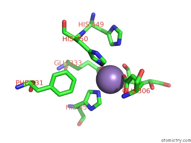
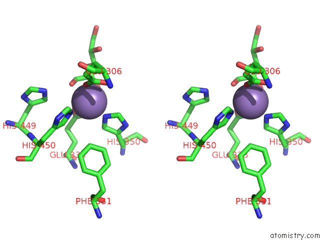
Manganese binding site 1 out of 3 in 4f2d
Go back to
Manganese binding site 1 out
of 3 in the Crystal Structure of Escherichia Coli L-Arabinose Isomerase (Ecai) Complexed with Ribitol
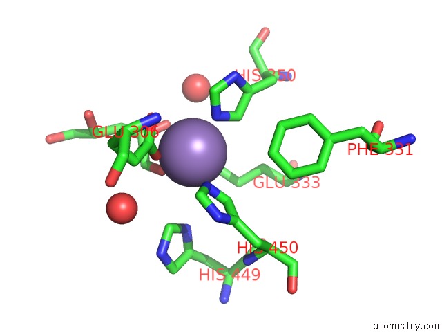
Mono view
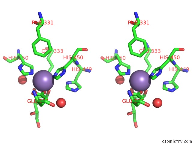
Stereo pair view
Mono view
Stereo pair view
A full contact list of Manganese with other atoms in the Mn binding
site number 1 of Crystal Structure of Escherichia Coli L-Arabinose Isomerase (Ecai) Complexed with Ribitol within 5.0Å range:
|
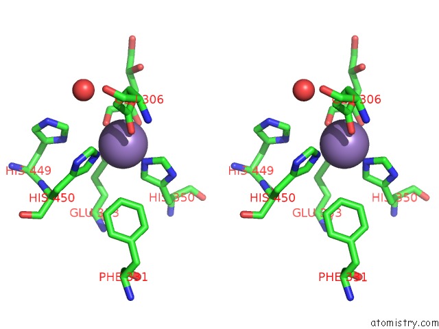
Manganese binding site 2 out of 3 in 4f2d
Go back to
Manganese binding site 2 out
of 3 in the Crystal Structure of Escherichia Coli L-Arabinose Isomerase (Ecai) Complexed with Ribitol
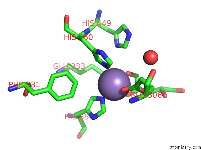
Mono view
Stereo pair view
Mono view
Stereo pair view
A full contact list of Manganese with other atoms in the Mn binding
site number 2 of Crystal Structure of Escherichia Coli L-Arabinose Isomerase (Ecai) Complexed with Ribitol within 5.0Å range:
|
Manganese binding site 3 out of 3 in 4f2d
Go back to
Manganese binding site 3 out
of 3 in the Crystal Structure of Escherichia Coli L-Arabinose Isomerase (Ecai) Complexed with Ribitol
Mono view
Stereo pair view
Mono view
Stereo pair view
A full contact list of Manganese with other atoms in the Mn binding
site number 3 of Crystal Structure of Escherichia Coli L-Arabinose Isomerase (Ecai) Complexed with Ribitol within 5.0Å range:
|
Reference:
B.A.Manjasetty,
S.K.Burley,
S.C.Almo,
M.R.Chance.
Crystal Structure of Escherichia Coli L-Arabinose Isomerase (Ecai) Complexed with Ribitol To Be Published.
Page generated: Sat Oct 5 19:20:58 2024
Last articles
K in 6CP4K in 6CNN
K in 6CNM
K in 6CK5
K in 6CGP
K in 6CF1
K in 6CI0
K in 6C9X
K in 6C65
K in 6C64