Manganese »
PDB 4ee3-4fo6 »
4ewt »
Manganese in PDB 4ewt: The Crystal Structure of A Putative Aminohydrolase From Methicillin Resistant Staphylococcus Aureus
Enzymatic activity of The Crystal Structure of A Putative Aminohydrolase From Methicillin Resistant Staphylococcus Aureus
All present enzymatic activity of The Crystal Structure of A Putative Aminohydrolase From Methicillin Resistant Staphylococcus Aureus:
3.5.1.14;
3.5.1.14;
Protein crystallography data
The structure of The Crystal Structure of A Putative Aminohydrolase From Methicillin Resistant Staphylococcus Aureus, PDB code: 4ewt
was solved by
T.S.Girish,
B.Vivek,
M.Colaco,
S.Misquith,
B.Gopal,
with X-Ray Crystallography technique. A brief refinement statistics is given in the table below:
| Resolution Low / High (Å) | 39.32 / 2.10 |
| Space group | P 1 |
| Cell size a, b, c (Å), α, β, γ (°) | 44.620, 120.110, 132.410, 115.40, 94.64, 96.55 |
| R / Rfree (%) | 19.9 / 22.9 |
Manganese Binding Sites:
The binding sites of Manganese atom in the The Crystal Structure of A Putative Aminohydrolase From Methicillin Resistant Staphylococcus Aureus
(pdb code 4ewt). This binding sites where shown within
5.0 Angstroms radius around Manganese atom.
In total 8 binding sites of Manganese where determined in the The Crystal Structure of A Putative Aminohydrolase From Methicillin Resistant Staphylococcus Aureus, PDB code: 4ewt:
Jump to Manganese binding site number: 1; 2; 3; 4; 5; 6; 7; 8;
In total 8 binding sites of Manganese where determined in the The Crystal Structure of A Putative Aminohydrolase From Methicillin Resistant Staphylococcus Aureus, PDB code: 4ewt:
Jump to Manganese binding site number: 1; 2; 3; 4; 5; 6; 7; 8;
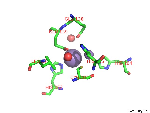
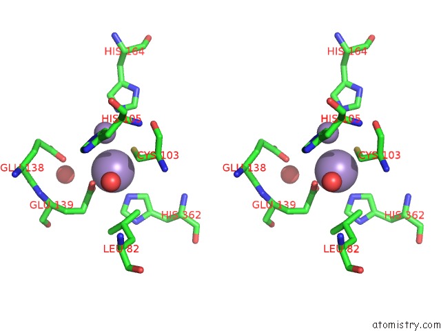
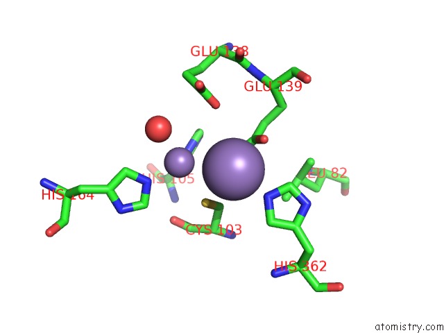
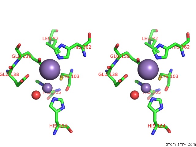
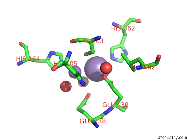
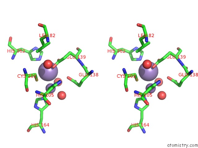
Manganese binding site 1 out of 8 in 4ewt
Go back to
Manganese binding site 1 out
of 8 in the The Crystal Structure of A Putative Aminohydrolase From Methicillin Resistant Staphylococcus Aureus
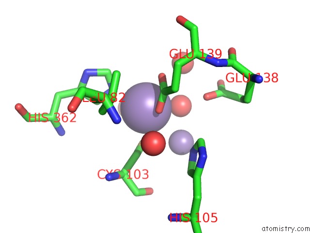
Mono view
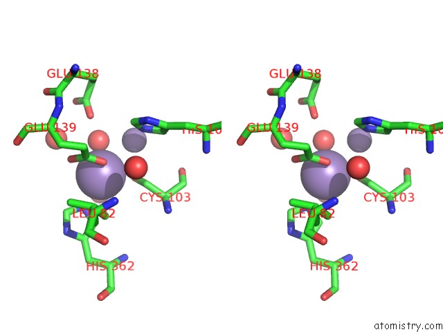
Stereo pair view
Mono view
Stereo pair view
A full contact list of Manganese with other atoms in the Mn binding
site number 1 of The Crystal Structure of A Putative Aminohydrolase From Methicillin Resistant Staphylococcus Aureus within 5.0Å range:
|
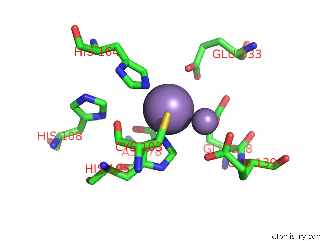
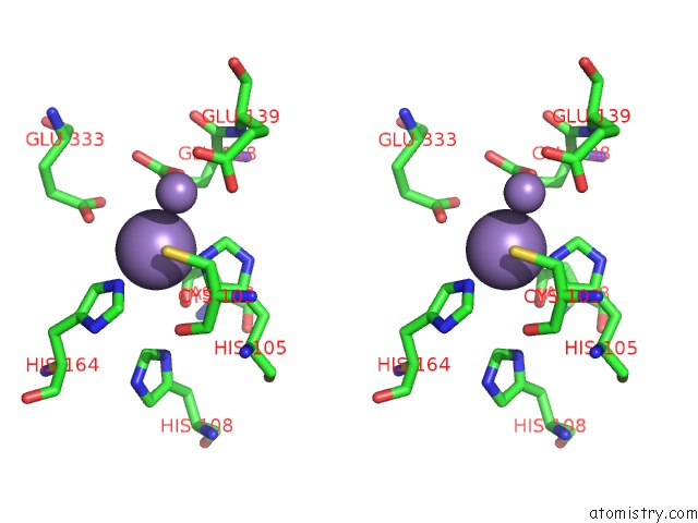
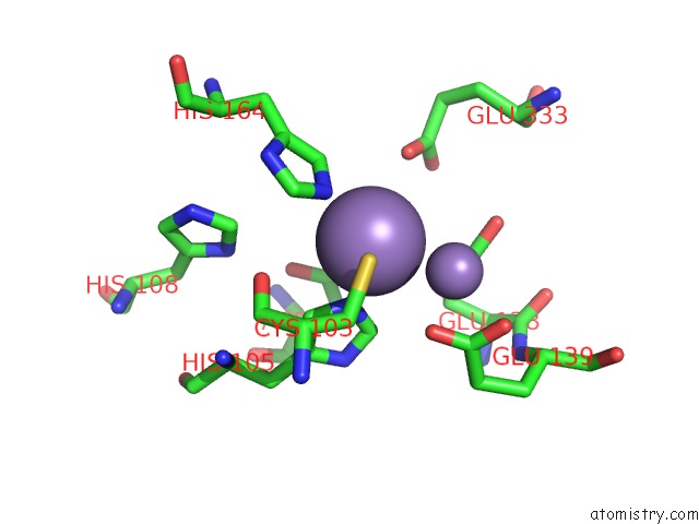
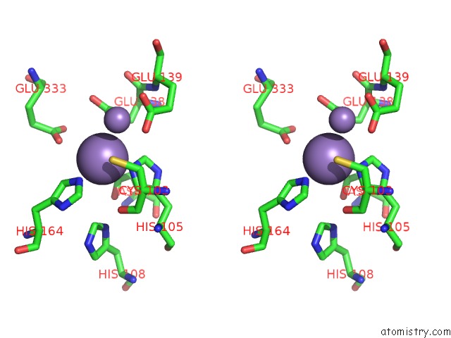
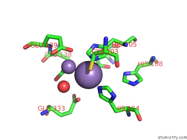
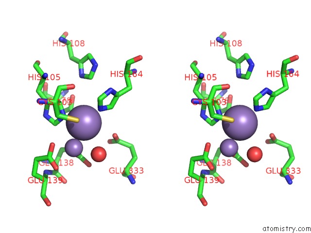
Manganese binding site 2 out of 8 in 4ewt
Go back to
Manganese binding site 2 out
of 8 in the The Crystal Structure of A Putative Aminohydrolase From Methicillin Resistant Staphylococcus Aureus
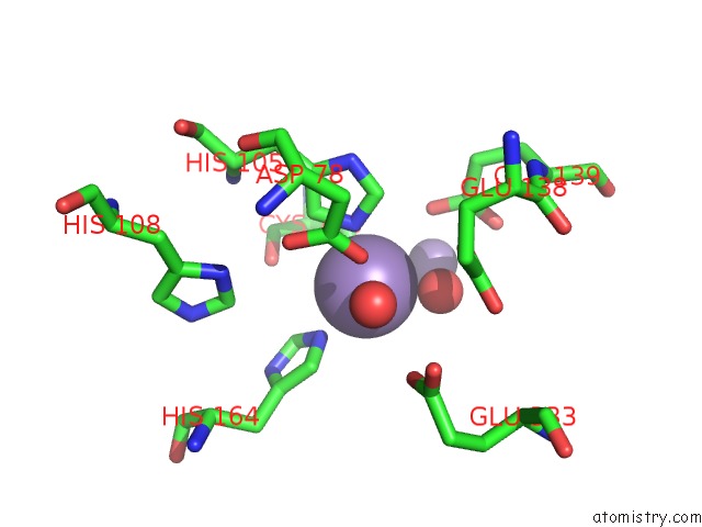
Mono view
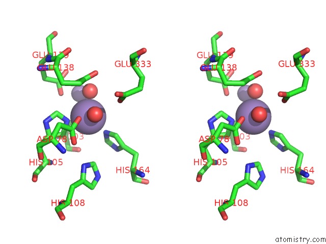
Stereo pair view
Mono view
Stereo pair view
A full contact list of Manganese with other atoms in the Mn binding
site number 2 of The Crystal Structure of A Putative Aminohydrolase From Methicillin Resistant Staphylococcus Aureus within 5.0Å range:
|
Manganese binding site 3 out of 8 in 4ewt
Go back to
Manganese binding site 3 out
of 8 in the The Crystal Structure of A Putative Aminohydrolase From Methicillin Resistant Staphylococcus Aureus
Mono view
Stereo pair view
Mono view
Stereo pair view
A full contact list of Manganese with other atoms in the Mn binding
site number 3 of The Crystal Structure of A Putative Aminohydrolase From Methicillin Resistant Staphylococcus Aureus within 5.0Å range:
|
Manganese binding site 4 out of 8 in 4ewt
Go back to
Manganese binding site 4 out
of 8 in the The Crystal Structure of A Putative Aminohydrolase From Methicillin Resistant Staphylococcus Aureus
Mono view
Stereo pair view
Mono view
Stereo pair view
A full contact list of Manganese with other atoms in the Mn binding
site number 4 of The Crystal Structure of A Putative Aminohydrolase From Methicillin Resistant Staphylococcus Aureus within 5.0Å range:
|
Manganese binding site 5 out of 8 in 4ewt
Go back to
Manganese binding site 5 out
of 8 in the The Crystal Structure of A Putative Aminohydrolase From Methicillin Resistant Staphylococcus Aureus
Mono view
Stereo pair view
Mono view
Stereo pair view
A full contact list of Manganese with other atoms in the Mn binding
site number 5 of The Crystal Structure of A Putative Aminohydrolase From Methicillin Resistant Staphylococcus Aureus within 5.0Å range:
|
Manganese binding site 6 out of 8 in 4ewt
Go back to
Manganese binding site 6 out
of 8 in the The Crystal Structure of A Putative Aminohydrolase From Methicillin Resistant Staphylococcus Aureus
Mono view
Stereo pair view
Mono view
Stereo pair view
A full contact list of Manganese with other atoms in the Mn binding
site number 6 of The Crystal Structure of A Putative Aminohydrolase From Methicillin Resistant Staphylococcus Aureus within 5.0Å range:
|
Manganese binding site 7 out of 8 in 4ewt
Go back to
Manganese binding site 7 out
of 8 in the The Crystal Structure of A Putative Aminohydrolase From Methicillin Resistant Staphylococcus Aureus
Mono view
Stereo pair view
Mono view
Stereo pair view
A full contact list of Manganese with other atoms in the Mn binding
site number 7 of The Crystal Structure of A Putative Aminohydrolase From Methicillin Resistant Staphylococcus Aureus within 5.0Å range:
|
Manganese binding site 8 out of 8 in 4ewt
Go back to
Manganese binding site 8 out
of 8 in the The Crystal Structure of A Putative Aminohydrolase From Methicillin Resistant Staphylococcus Aureus
Mono view
Stereo pair view
Mono view
Stereo pair view
A full contact list of Manganese with other atoms in the Mn binding
site number 8 of The Crystal Structure of A Putative Aminohydrolase From Methicillin Resistant Staphylococcus Aureus within 5.0Å range:
|
Reference:
T.S.Girish,
B.Vivek,
M.Colaco,
S.Misquith,
B.Gopal.
Structure of An Amidohydrolase, SACOL0085, From Methicillin-Resistant Staphylococcus Aureus Col Acta Crystallogr.,Sect.F V. 69 103 2013.
ISSN: ESSN 1744-3091
PubMed: 23385746
DOI: 10.1107/S1744309112049822
Page generated: Sat Oct 5 19:20:52 2024
ISSN: ESSN 1744-3091
PubMed: 23385746
DOI: 10.1107/S1744309112049822
Last articles
K in 2YNMK in 2YI8
K in 2YIF
K in 2YIE
K in 2YHK
K in 2YCP
K in 2YGH
K in 2YD0
K in 2YFY
K in 2YCT