Manganese »
PDB 4b5i-4dec »
4ccl »
Manganese in PDB 4ccl: X-Ray Structure of E. Coli Ycfd
Protein crystallography data
The structure of X-Ray Structure of E. Coli Ycfd, PDB code: 4ccl
was solved by
M.A.Mcdonough,
C.H.Ho,
N.J.Kershaw,
C.J.Schofield,
with X-Ray Crystallography technique. A brief refinement statistics is given in the table below:
| Resolution Low / High (Å) | 41.765 / 2.60 |
| Space group | P 43 21 2 |
| Cell size a, b, c (Å), α, β, γ (°) | 120.693, 120.693, 133.498, 90.00, 90.00, 90.00 |
| R / Rfree (%) | 19.83 / 24.9 |
Manganese Binding Sites:
The binding sites of Manganese atom in the X-Ray Structure of E. Coli Ycfd
(pdb code 4ccl). This binding sites where shown within
5.0 Angstroms radius around Manganese atom.
In total 4 binding sites of Manganese where determined in the X-Ray Structure of E. Coli Ycfd, PDB code: 4ccl:
Jump to Manganese binding site number: 1; 2; 3; 4;
In total 4 binding sites of Manganese where determined in the X-Ray Structure of E. Coli Ycfd, PDB code: 4ccl:
Jump to Manganese binding site number: 1; 2; 3; 4;
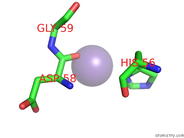
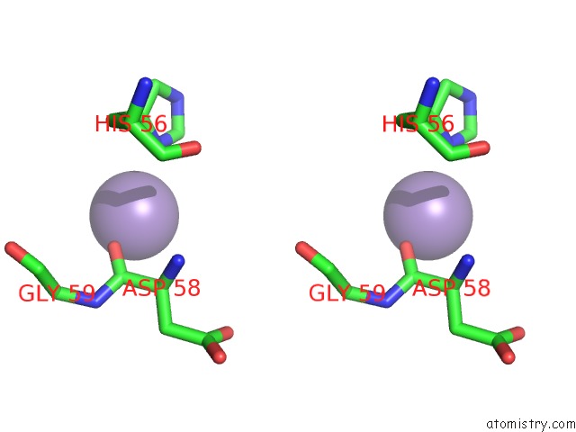
Manganese binding site 1 out of 4 in 4ccl
Go back to
Manganese binding site 1 out
of 4 in the X-Ray Structure of E. Coli Ycfd
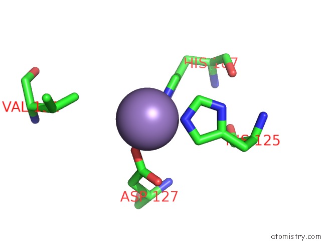
Mono view
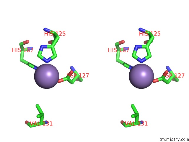
Stereo pair view
Mono view
Stereo pair view
A full contact list of Manganese with other atoms in the Mn binding
site number 1 of X-Ray Structure of E. Coli Ycfd within 5.0Å range:
|
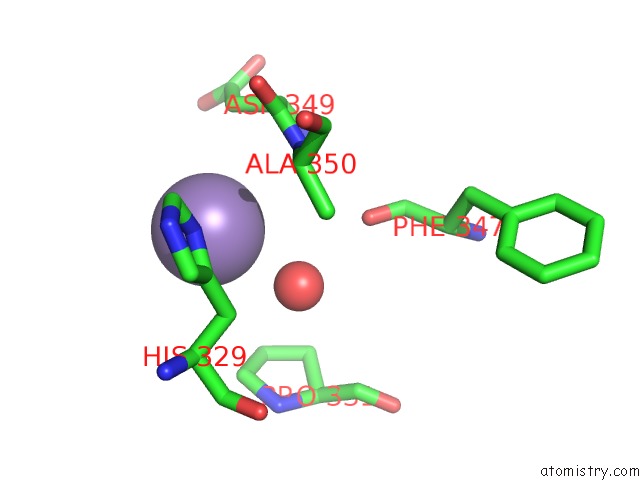
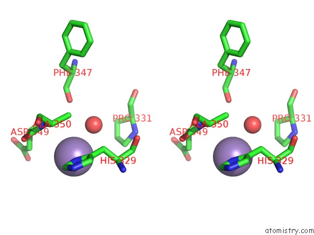
Manganese binding site 2 out of 4 in 4ccl
Go back to
Manganese binding site 2 out
of 4 in the X-Ray Structure of E. Coli Ycfd
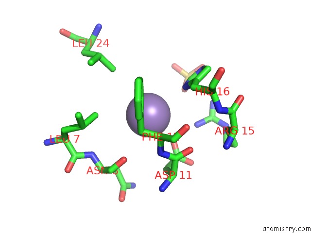
Mono view
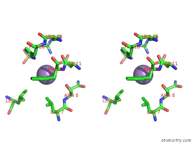
Stereo pair view
Mono view
Stereo pair view
A full contact list of Manganese with other atoms in the Mn binding
site number 2 of X-Ray Structure of E. Coli Ycfd within 5.0Å range:
|
Manganese binding site 3 out of 4 in 4ccl
Go back to
Manganese binding site 3 out
of 4 in the X-Ray Structure of E. Coli Ycfd
Mono view
Stereo pair view
Mono view
Stereo pair view
A full contact list of Manganese with other atoms in the Mn binding
site number 3 of X-Ray Structure of E. Coli Ycfd within 5.0Å range:
|
Manganese binding site 4 out of 4 in 4ccl
Go back to
Manganese binding site 4 out
of 4 in the X-Ray Structure of E. Coli Ycfd
Mono view
Stereo pair view
Mono view
Stereo pair view
A full contact list of Manganese with other atoms in the Mn binding
site number 4 of X-Ray Structure of E. Coli Ycfd within 5.0Å range:
|
Reference:
R.Chowdhury,
R.Sekirnik,
N.C.Brissett,
T.Krojer,
C.-H.Ho,
S.S.Ng,
I.J.Clifton,
W.Ge,
N.J.Kershaw,
G.C.Fox,
J.R.C.Muniz,
M.Vollmar,
C.Phillips,
E.S.Pilka,
K.L.Kavanagh,
F.Von Deflt,
U.Oppermann,
M.A.Mcdonough,
A.J.Doherty,
C.J.Schofield.
Ribosomal Oxygenases Are Structurally Conserved From Prokaryotes to Humans. Nature V. 510 422 2014.
ISSN: ISSN 0028-0836
PubMed: 24814345
DOI: 10.1038/NATURE13263
Page generated: Sat Oct 5 18:54:03 2024
ISSN: ISSN 0028-0836
PubMed: 24814345
DOI: 10.1038/NATURE13263
Last articles
Mg in 4DCAMg in 4DBR
Mg in 4DBH
Mg in 4DB8
Mg in 4DBQ
Mg in 4DBF
Mg in 4DA1
Mg in 4D6O
Mg in 4DAT
Mg in 4D6N