Manganese »
PDB 3vrs-3x2x »
3wak »
Manganese in PDB 3wak: Crystal Structure of the Archaeoglobus Fulgidus Oligosaccharyltransferase (O29867_ARCFU) in the Apo Form
Enzymatic activity of Crystal Structure of the Archaeoglobus Fulgidus Oligosaccharyltransferase (O29867_ARCFU) in the Apo Form
All present enzymatic activity of Crystal Structure of the Archaeoglobus Fulgidus Oligosaccharyltransferase (O29867_ARCFU) in the Apo Form:
2.4.1.119;
2.4.1.119;
Protein crystallography data
The structure of Crystal Structure of the Archaeoglobus Fulgidus Oligosaccharyltransferase (O29867_ARCFU) in the Apo Form, PDB code: 3wak
was solved by
S.Matsumoto,
A.Shimada,
D.Kohda,
with X-Ray Crystallography technique. A brief refinement statistics is given in the table below:
| Resolution Low / High (Å) | 49.90 / 3.41 |
| Space group | P 43 21 2 |
| Cell size a, b, c (Å), α, β, γ (°) | 123.357, 123.357, 182.496, 90.00, 90.00, 90.00 |
| R / Rfree (%) | 21.9 / 27.2 |
Manganese Binding Sites:
The binding sites of Manganese atom in the Crystal Structure of the Archaeoglobus Fulgidus Oligosaccharyltransferase (O29867_ARCFU) in the Apo Form
(pdb code 3wak). This binding sites where shown within
5.0 Angstroms radius around Manganese atom.
In total only one binding site of Manganese was determined in the Crystal Structure of the Archaeoglobus Fulgidus Oligosaccharyltransferase (O29867_ARCFU) in the Apo Form, PDB code: 3wak:
In total only one binding site of Manganese was determined in the Crystal Structure of the Archaeoglobus Fulgidus Oligosaccharyltransferase (O29867_ARCFU) in the Apo Form, PDB code: 3wak:
Manganese binding site 1 out of 1 in 3wak
Go back to
Manganese binding site 1 out
of 1 in the Crystal Structure of the Archaeoglobus Fulgidus Oligosaccharyltransferase (O29867_ARCFU) in the Apo Form
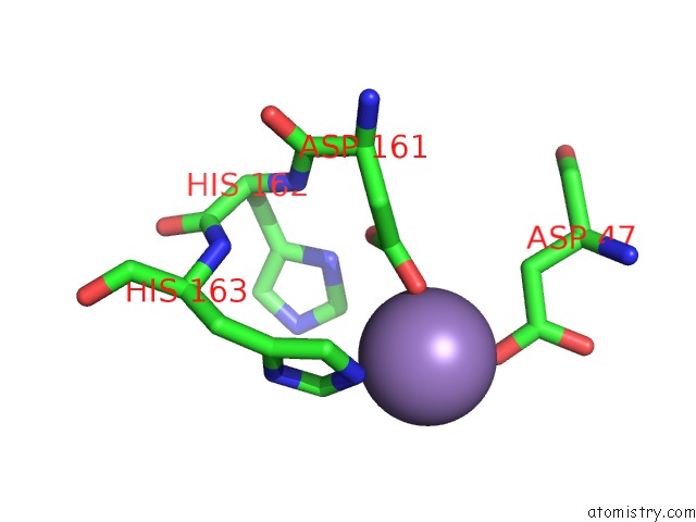
Mono view
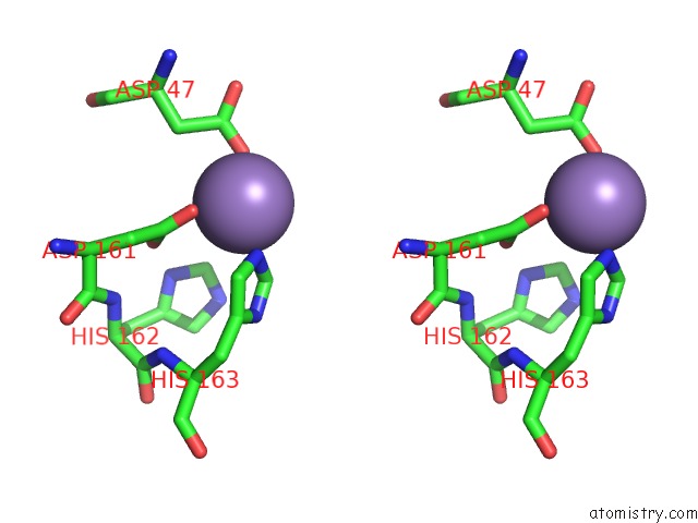
Stereo pair view
Mono view
Stereo pair view
A full contact list of Manganese with other atoms in the Mn binding
site number 1 of Crystal Structure of the Archaeoglobus Fulgidus Oligosaccharyltransferase (O29867_ARCFU) in the Apo Form within 5.0Å range:
|
Reference:
S.Matsumoto,
A.Shimada,
J.Nyirenda,
M.Igura,
Y.Kawano,
D.Kohda.
Crystal Structures of An Archaeal Oligosaccharyltransferase Provide Insights Into the Catalytic Cycle of N-Linked Protein Glycosylation Proc.Natl.Acad.Sci.Usa V. 110 17868 2013.
ISSN: ISSN 0027-8424
PubMed: 24127570
DOI: 10.1073/PNAS.1309777110
Page generated: Sat Aug 16 13:17:49 2025
ISSN: ISSN 0027-8424
PubMed: 24127570
DOI: 10.1073/PNAS.1309777110
Last articles
Br in 7HJ2Br in 7HJI
As in 9NBW
As in 9NBO
As in 9N0J
Mn in 9LJU
Mn in 9LJW
Mn in 9LJS
Mn in 9LJR
Mn in 9LJT