Atomistry » Manganese » PDB 3uag-3vnm » 3uu0
Atomistry »
  Manganese »
    PDB 3uag-3vnm »
      3uu0 »

Manganese in PDB 3uu0: Crystal Structure of L-Rhamnose Isomerase From Bacillus Halodurans in Complex with Mn

Enzymatic activity of Crystal Structure of L-Rhamnose Isomerase From Bacillus Halodurans in Complex with Mn

All present enzymatic activity of Crystal Structure of L-Rhamnose Isomerase From Bacillus Halodurans in Complex with Mn:
5.3.1.14;

Protein crystallography data

The structure of Crystal Structure of L-Rhamnose Isomerase From Bacillus Halodurans in Complex with Mn, PDB code: 3uu0 was solved by T.T.N.Doan, P.Prabhu, J.K.Kim, M.Jeya, L.W.Kang, J.K.Lee, with X-Ray Crystallography technique. A brief refinement statistics is given in the table below:

Resolution Low / High (Å) 46.62 / 2.70
Space group P 1 21 1
Cell size a, b, c (Å), α, β, γ (°) 83.584, 165.553, 92.471, 90.00, 115.82, 90.00
R / Rfree (%) 18 / 25.5

Manganese Binding Sites:

The binding sites of Manganese atom in the Crystal Structure of L-Rhamnose Isomerase From Bacillus Halodurans in Complex with Mn (pdb code 3uu0). This binding sites where shown within 5.0 Angstroms radius around Manganese atom.
In total 8 binding sites of Manganese where determined in the Crystal Structure of L-Rhamnose Isomerase From Bacillus Halodurans in Complex with Mn, PDB code: 3uu0:
Jump to Manganese binding site number: 1; 2; 3; 4; 5; 6; 7; 8;

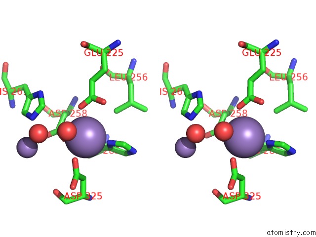
Manganese binding site 1 out of 8 in 3uu0

Go back to Manganese Binding Sites List in 3uu0
Manganese binding site 1 out of 8 in the Crystal Structure of L-Rhamnose Isomerase From Bacillus Halodurans in Complex with Mn


Mono view


Stereo pair view

A full contact list of Manganese with other atoms in the Mn binding site number 1 of Crystal Structure of L-Rhamnose Isomerase From Bacillus Halodurans in Complex with Mn within 5.0Å range:
probe atom residue distance (Å) B Occ
A:Mn419

b:45.8
occ:1.00
OD2 A:ASP258 2.0 29.0 1.0
OD2 A:ASP325 2.1 36.8 1.0
OE2 A:GLU225 2.2 27.7 1.0
ND1 A:HIS285 2.3 31.5 1.0
O A:HOH472 2.4 21.6 1.0
O A:HOH473 2.5 34.5 1.0
CG A:ASP258 3.1 25.5 1.0
CD A:GLU225 3.1 23.2 1.0
CE1 A:HIS285 3.2 32.0 1.0
CG A:ASP325 3.2 31.6 1.0
CG A:HIS285 3.3 29.1 1.0
OE1 A:GLU225 3.5 22.9 1.0
CB A:HIS285 3.6 28.1 1.0
CB A:ASP258 3.7 23.4 1.0
CB A:ASP325 3.8 30.2 1.0
CE1 A:HIS261 4.1 26.1 1.0
OD1 A:ASP258 4.2 27.4 1.0
OD1 A:ASP325 4.3 33.1 1.0
NE2 A:HIS285 4.3 31.5 1.0
CD2 A:HIS285 4.4 28.6 1.0
NE2 A:HIS261 4.4 26.6 1.0
CG A:GLU225 4.4 21.6 1.0
MN A:MN420 4.5 51.5 1.0
CD2 A:LEU256 4.8 21.9 1.0
ND1 A:HIS261 4.8 27.9 1.0
CA A:ASP258 4.8 23.6 1.0

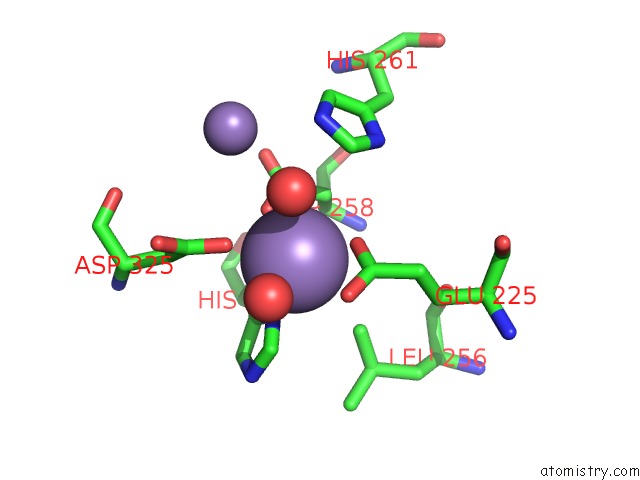
Manganese binding site 2 out of 8 in 3uu0

Go back to Manganese Binding Sites List in 3uu0
Manganese binding site 2 out of 8 in the Crystal Structure of L-Rhamnose Isomerase From Bacillus Halodurans in Complex with Mn


Mono view


Stereo pair view

A full contact list of Manganese with other atoms in the Mn binding site number 2 of Crystal Structure of L-Rhamnose Isomerase From Bacillus Halodurans in Complex with Mn within 5.0Å range:
probe atom residue distance (Å) B Occ
A:Mn420

b:51.5
occ:1.00
OD1 A:ASP293 1.9 28.6 1.0
OD2 A:ASP293 2.2 27.7 1.0
NE2 A:HIS261 2.3 26.6 1.0
CG A:ASP293 2.4 25.3 1.0
OD2 A:ASP295 2.9 36.0 1.0
CD2 A:HIS261 2.9 25.3 1.0
O A:HOH472 3.1 21.6 1.0
OD1 A:ASP258 3.4 27.4 1.0
CE1 A:HIS261 3.4 26.1 1.0
CG A:ASP295 3.7 32.1 1.0
OD1 A:ASP295 3.7 33.9 1.0
OD2 A:ASP258 3.7 29.0 1.0
CG A:ASP258 3.8 25.5 1.0
CB A:ASP293 3.9 22.5 1.0
O A:HOH469 4.1 35.5 1.0
CG A:HIS261 4.2 25.8 1.0
ND1 A:HIS261 4.3 27.9 1.0
OG A:SER287 4.4 25.7 1.0
MN A:MN419 4.5 45.8 1.0
NZ A:LYS227 4.6 27.8 1.0
OD2 A:ASP325 4.6 36.8 1.0
O A:ASP325 4.8 32.5 1.0
CA A:ASP293 4.8 23.7 1.0
CA A:GLY260 4.9 23.6 1.0
CB A:ASP258 4.9 23.4 1.0
CG A:ASP325 4.9 31.6 1.0
C A:GLY260 4.9 22.9 1.0
O A:GLY260 5.0 24.2 1.0

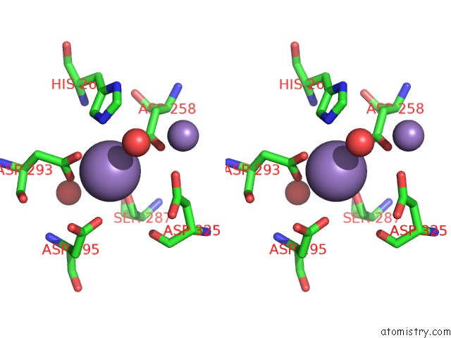
Manganese binding site 3 out of 8 in 3uu0

Go back to Manganese Binding Sites List in 3uu0
Manganese binding site 3 out of 8 in the Crystal Structure of L-Rhamnose Isomerase From Bacillus Halodurans in Complex with Mn


Mono view


Stereo pair view

A full contact list of Manganese with other atoms in the Mn binding site number 3 of Crystal Structure of L-Rhamnose Isomerase From Bacillus Halodurans in Complex with Mn within 5.0Å range:
probe atom residue distance (Å) B Occ
B:Mn419

b:37.3
occ:1.00
OD2 B:ASP325 2.3 35.4 1.0
ND1 B:HIS285 2.3 32.2 1.0
OD2 B:ASP258 2.3 24.7 1.0
OE2 B:GLU225 2.6 24.8 1.0
CG B:ASP325 3.2 29.1 1.0
CG B:HIS285 3.3 30.2 1.0
CE1 B:HIS285 3.3 33.1 1.0
CG B:ASP258 3.4 21.2 1.0
CD B:GLU225 3.4 24.4 1.0
CB B:ASP325 3.5 27.4 1.0
CB B:HIS285 3.5 27.0 1.0
OE1 B:GLU225 3.5 24.9 1.0
CB B:ASP258 3.8 20.6 1.0
O B:HOH471 4.1 37.8 1.0
CD1 B:LEU256 4.3 20.9 1.0
OD1 B:ASP325 4.4 27.2 1.0
NE2 B:HIS285 4.4 32.7 1.0
CE1 B:HIS261 4.4 21.5 1.0
CD2 B:HIS285 4.4 30.4 1.0
OD1 B:ASP258 4.5 20.4 1.0
O B:HOH477 4.6 43.7 1.0
NE2 B:HIS261 4.7 19.9 1.0
CG B:GLU225 4.7 24.3 1.0
MN B:MN420 4.9 63.7 1.0
CA B:ASP258 5.0 20.9 1.0
CA B:ASP325 5.0 28.0 1.0

Manganese binding site 4 out of 8 in 3uu0

Go back to Manganese Binding Sites List in 3uu0
Manganese binding site 4 out of 8 in the Crystal Structure of L-Rhamnose Isomerase From Bacillus Halodurans in Complex with Mn


Mono view


Stereo pair view

A full contact list of Manganese with other atoms in the Mn binding site number 4 of Crystal Structure of L-Rhamnose Isomerase From Bacillus Halodurans in Complex with Mn within 5.0Å range:
probe atom residue distance (Å) B Occ
B:Mn420

b:63.7
occ:1.00
OD2 B:ASP293 2.3 31.3 1.0
O B:HOH477 2.4 43.7 1.0
NE2 B:HIS261 2.6 19.9 1.0
OD1 B:ASP293 2.8 23.4 1.0
CG B:ASP293 2.9 25.3 1.0
OD1 B:ASP295 3.2 34.8 1.0
CD2 B:HIS261 3.4 19.0 1.0
CE1 B:HIS261 3.6 21.5 1.0
OD2 B:ASP258 3.9 24.7 1.0
CG B:ASP295 3.9 30.0 1.0
OD2 B:ASP295 4.0 35.0 1.0
OD2 B:ASP325 4.0 35.4 1.0
OD1 B:ASP258 4.3 20.4 1.0
CG B:ASP258 4.3 21.2 1.0
CB B:ASP293 4.3 22.5 1.0
O B:HOH471 4.5 37.8 1.0
CG B:ASP325 4.6 29.1 1.0
CG B:HIS261 4.6 20.0 1.0
NZ B:LYS227 4.7 28.3 1.0
O B:HOH442 4.7 17.4 1.0
ND1 B:HIS261 4.7 20.7 1.0
OG B:SER287 4.9 25.8 1.0
OD1 B:ASP325 4.9 27.2 1.0
MN B:MN419 4.9 37.3 1.0
O B:ASP293 5.0 22.1 1.0
O B:ASP325 5.0 31.6 1.0

Manganese binding site 5 out of 8 in 3uu0

Go back to Manganese Binding Sites List in 3uu0
Manganese binding site 5 out of 8 in the Crystal Structure of L-Rhamnose Isomerase From Bacillus Halodurans in Complex with Mn


Mono view


Stereo pair view

A full contact list of Manganese with other atoms in the Mn binding site number 5 of Crystal Structure of L-Rhamnose Isomerase From Bacillus Halodurans in Complex with Mn within 5.0Å range:
probe atom residue distance (Å) B Occ
C:Mn419

b:44.6
occ:1.00
OE1 C:GLU225 2.1 32.6 1.0
OD2 C:ASP325 2.3 35.9 1.0
OE2 C:GLU225 2.3 29.8 1.0
ND1 C:HIS285 2.4 28.8 1.0
O C:HOH476 2.5 26.8 1.0
CD C:GLU225 2.5 29.4 1.0
OD2 C:ASP258 2.6 30.9 1.0
CE1 C:HIS285 3.1 27.9 1.0
O C:HOH475 3.2 36.7 1.0
CG C:ASP325 3.5 31.4 1.0
CG C:HIS285 3.6 27.0 1.0
CG C:ASP258 3.6 22.9 1.0
CB C:ASP258 3.9 19.7 1.0
CG C:GLU225 4.0 25.7 1.0
CB C:HIS285 4.0 28.0 1.0
CE1 C:HIS261 4.1 28.8 1.0
CD2 C:LEU256 4.2 24.1 1.0
NE2 C:HIS285 4.3 26.3 1.0
OD1 C:ASP325 4.4 35.6 1.0
CB C:ASP325 4.4 27.9 1.0
CD2 C:HIS285 4.6 24.6 1.0
NE2 C:HIS261 4.6 28.9 1.0
MN C:MN420 4.7 53.3 1.0
OD1 C:ASP258 4.7 23.1 1.0
ND1 C:HIS261 4.8 28.6 1.0
CB C:GLU225 4.8 24.9 1.0

Manganese binding site 6 out of 8 in 3uu0

Go back to Manganese Binding Sites List in 3uu0
Manganese binding site 6 out of 8 in the Crystal Structure of L-Rhamnose Isomerase From Bacillus Halodurans in Complex with Mn


Mono view


Stereo pair view

A full contact list of Manganese with other atoms in the Mn binding site number 6 of Crystal Structure of L-Rhamnose Isomerase From Bacillus Halodurans in Complex with Mn within 5.0Å range:
probe atom residue distance (Å) B Occ
C:Mn420

b:53.3
occ:1.00
OD2 C:ASP293 2.0 31.1 1.0
CG C:ASP293 2.8 28.5 1.0
OD1 C:ASP293 2.8 28.3 1.0
NE2 C:HIS261 2.9 28.9 1.0
OD1 C:ASP295 3.0 37.4 1.0
OD1 C:ASP258 3.2 23.1 1.0
OD2 C:ASP258 3.3 30.9 1.0
CD2 C:HIS261 3.5 26.5 1.0
CG C:ASP258 3.5 22.9 1.0
OD2 C:ASP325 3.7 35.9 1.0
O C:HOH475 3.8 36.7 1.0
CG C:ASP295 3.8 32.4 1.0
OD2 C:ASP295 3.9 33.3 1.0
CE1 C:HIS261 3.9 28.8 1.0
CG C:ASP325 4.1 31.4 1.0
OG C:SER287 4.2 33.0 1.0
CB C:ASP293 4.3 26.3 1.0
OD1 C:ASP325 4.4 35.6 1.0
CG C:HIS261 4.6 25.7 1.0
MN C:MN419 4.7 44.6 1.0
O C:ASP325 4.7 29.2 1.0
O C:HOH434 4.8 32.3 1.0
CB C:ASP325 4.8 27.9 1.0
ND1 C:HIS261 4.8 28.6 1.0
CB C:ASP258 4.8 19.7 1.0
CB C:SER287 5.0 31.2 1.0

Manganese binding site 7 out of 8 in 3uu0

Go back to Manganese Binding Sites List in 3uu0
Manganese binding site 7 out of 8 in the Crystal Structure of L-Rhamnose Isomerase From Bacillus Halodurans in Complex with Mn


Mono view


Stereo pair view

A full contact list of Manganese with other atoms in the Mn binding site number 7 of Crystal Structure of L-Rhamnose Isomerase From Bacillus Halodurans in Complex with Mn within 5.0Å range:
probe atom residue distance (Å) B Occ
D:Mn419

b:50.8
occ:1.00
OD1 D:ASP258 2.0 27.2 1.0
O D:HOH478 2.3 30.0 1.0
ND1 D:HIS285 2.3 33.2 1.0
OE2 D:GLU225 2.4 32.2 1.0
OD1 D:ASP325 2.4 39.9 1.0
O D:HOH477 2.5 30.0 1.0
CD D:GLU225 3.2 29.0 1.0
CG D:ASP325 3.3 40.7 1.0
CG D:ASP258 3.3 26.1 1.0
CE1 D:HIS285 3.3 36.4 1.0
CG D:HIS285 3.3 31.6 1.0
OE1 D:GLU225 3.5 30.0 1.0
CB D:HIS285 3.6 30.5 1.0
CB D:ASP325 3.7 39.9 1.0
OD2 D:ASP258 4.1 29.0 1.0
CB D:ASP258 4.1 24.1 1.0
OD2 D:ASP325 4.2 45.5 1.0
CD1 D:LEU256 4.4 29.9 1.0
NE2 D:HIS285 4.4 36.6 1.0
CE1 D:HIS261 4.4 27.1 1.0
CD2 D:HIS285 4.5 34.1 1.0
CG D:GLU225 4.5 27.7 1.0
O D:HOH441 4.7 38.5 1.0
MN D:MN420 4.7 55.1 1.0
O D:HOH467 4.7 38.9 1.0
NE2 D:HIS261 4.8 28.3 1.0

Manganese binding site 8 out of 8 in 3uu0

Go back to Manganese Binding Sites List in 3uu0
Manganese binding site 8 out of 8 in the Crystal Structure of L-Rhamnose Isomerase From Bacillus Halodurans in Complex with Mn


Mono view


Stereo pair view

A full contact list of Manganese with other atoms in the Mn binding site number 8 of Crystal Structure of L-Rhamnose Isomerase From Bacillus Halodurans in Complex with Mn within 5.0Å range:
probe atom residue distance (Å) B Occ
D:Mn420

b:55.1
occ:1.00
OD2 D:ASP293 2.1 34.1 1.0
O D:HOH478 2.4 30.0 1.0
NE2 D:HIS261 2.4 28.3 1.0
OD1 D:ASP293 2.8 27.6 1.0
CG D:ASP293 2.8 29.1 1.0
O D:HOH467 2.9 38.9 1.0
OD1 D:ASP295 3.0 44.3 1.0
CD2 D:HIS261 3.4 26.1 1.0
CE1 D:HIS261 3.4 27.1 1.0
OD2 D:ASP295 3.5 41.7 1.0
CG D:ASP295 3.7 36.8 1.0
O D:HOH441 3.7 38.5 1.0
OD1 D:ASP325 3.9 39.9 1.0
OD1 D:ASP258 3.9 27.2 1.0
OD2 D:ASP258 4.1 29.0 1.0
CG D:ASP258 4.2 26.1 1.0
CB D:ASP293 4.3 27.2 1.0
NZ D:LYS227 4.4 28.3 1.0
ND1 D:HIS261 4.5 28.4 1.0
CG D:HIS261 4.5 25.9 1.0
O D:HOH477 4.6 30.0 1.0
MN D:MN419 4.7 50.8 1.0
CG D:ASP325 4.9 40.7 1.0
CD D:LYS227 4.9 27.0 1.0
OG D:SER287 5.0 30.0 1.0

Reference:

P.Prabhu, T.N.Doan, M.Tiwari, R.Singh, S.C.Kim, M.K.Hong, Y.C.Kang, L.W.Kang, J.K.Lee. Structure-Based Studies on the Metal Binding of Two-Metal-Dependent Sugar Isomerases. Febs J. V. 281 3446 2014.
ISSN: ISSN 1742-464X
PubMed: 24925069
DOI: 10.1111/FEBS.12872
Page generated: Sat Oct 5 18:14:20 2024

Last articles

K in 7QQP
K in 7QQO
K in 7QK5
K in 7QIX
K in 7QNO
K in 7QIY
K in 7Q3X
K in 7QDN
K in 7QF6
K in 7Q0G
© Copyright 2008-2020 by atomistry.com
Home   |    Site Map   |    Copyright   |    Contact us   |    Privacy