Manganese »
PDB 3uag-3vnm »
3ulq »
Manganese in PDB 3ulq: Crystal Structure of the Anti-Activator Rapf Complexed with the Response Regulator Coma Dna Binding Domain
Protein crystallography data
The structure of Crystal Structure of the Anti-Activator Rapf Complexed with the Response Regulator Coma Dna Binding Domain, PDB code: 3ulq
was solved by
M.D.Baker,
M.B.Neiditch,
with X-Ray Crystallography technique. A brief refinement statistics is given in the table below:
| Resolution Low / High (Å) | 19.76 / 2.30 |
| Space group | P 21 21 2 |
| Cell size a, b, c (Å), α, β, γ (°) | 115.304, 81.437, 78.346, 90.00, 90.00, 90.00 |
| R / Rfree (%) | 18.9 / 22.4 |
Manganese Binding Sites:
The binding sites of Manganese atom in the Crystal Structure of the Anti-Activator Rapf Complexed with the Response Regulator Coma Dna Binding Domain
(pdb code 3ulq). This binding sites where shown within
5.0 Angstroms radius around Manganese atom.
In total only one binding site of Manganese was determined in the Crystal Structure of the Anti-Activator Rapf Complexed with the Response Regulator Coma Dna Binding Domain, PDB code: 3ulq:
In total only one binding site of Manganese was determined in the Crystal Structure of the Anti-Activator Rapf Complexed with the Response Regulator Coma Dna Binding Domain, PDB code: 3ulq:
Manganese binding site 1 out of 1 in 3ulq
Go back to
Manganese binding site 1 out
of 1 in the Crystal Structure of the Anti-Activator Rapf Complexed with the Response Regulator Coma Dna Binding Domain
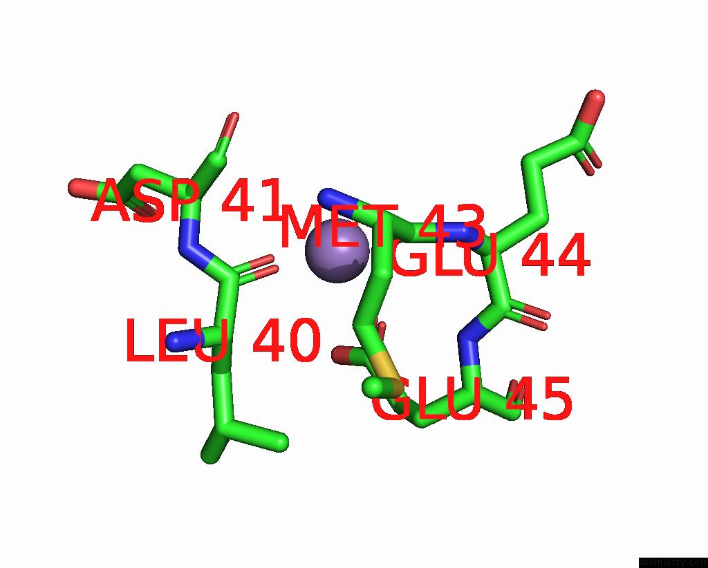
Mono view
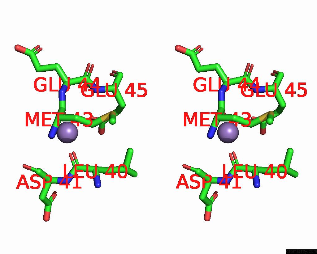
Stereo pair view
Mono view
Stereo pair view
A full contact list of Manganese with other atoms in the Mn binding
site number 1 of Crystal Structure of the Anti-Activator Rapf Complexed with the Response Regulator Coma Dna Binding Domain within 5.0Å range:
|
Reference:
M.D.Baker,
M.B.Neiditch.
Structural Basis of Response Regulator Inhibition By A Bacterial Anti-Activator Protein. Plos Biol. V. 9 01226 2011.
ISSN: ISSN 1544-9173
PubMed: 22215984
DOI: 10.1371/JOURNAL.PBIO.1001226
Page generated: Sat Oct 5 18:05:49 2024
ISSN: ISSN 1544-9173
PubMed: 22215984
DOI: 10.1371/JOURNAL.PBIO.1001226
Last articles
K in 6DP6K in 6DP4
K in 6DP5
K in 6DP7
K in 6DP3
K in 6DP2
K in 6DP1
K in 6DP0
K in 6DOR
K in 6DOS