Manganese »
PDB 3sx5-3u95 »
3tc3 »
Manganese in PDB 3tc3: Crystal Structure of Sacuvde
Protein crystallography data
The structure of Crystal Structure of Sacuvde, PDB code: 3tc3
was solved by
E.M.Meulenbroek,
I.Jala,
G.F.Moolenaar,
N.Goosen,
N.S.Pannu,
with X-Ray Crystallography technique. A brief refinement statistics is given in the table below:
| Resolution Low / High (Å) | 46.05 / 1.50 |
| Space group | P 1 |
| Cell size a, b, c (Å), α, β, γ (°) | 42.075, 53.585, 77.395, 102.10, 93.02, 111.77 |
| R / Rfree (%) | 17.7 / 21.4 |
Manganese Binding Sites:
The binding sites of Manganese atom in the Crystal Structure of Sacuvde
(pdb code 3tc3). This binding sites where shown within
5.0 Angstroms radius around Manganese atom.
In total 2 binding sites of Manganese where determined in the Crystal Structure of Sacuvde, PDB code: 3tc3:
Jump to Manganese binding site number: 1; 2;
In total 2 binding sites of Manganese where determined in the Crystal Structure of Sacuvde, PDB code: 3tc3:
Jump to Manganese binding site number: 1; 2;
Manganese binding site 1 out of 2 in 3tc3
Go back to
Manganese binding site 1 out
of 2 in the Crystal Structure of Sacuvde
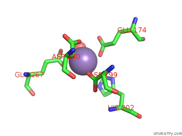
Mono view
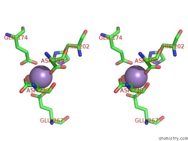
Stereo pair view
Mono view
Stereo pair view
A full contact list of Manganese with other atoms in the Mn binding
site number 1 of Crystal Structure of Sacuvde within 5.0Å range:
|
Manganese binding site 2 out of 2 in 3tc3
Go back to
Manganese binding site 2 out
of 2 in the Crystal Structure of Sacuvde
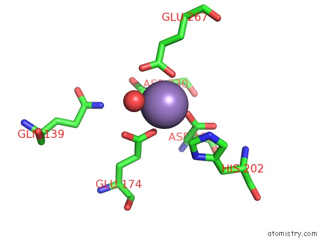
Mono view
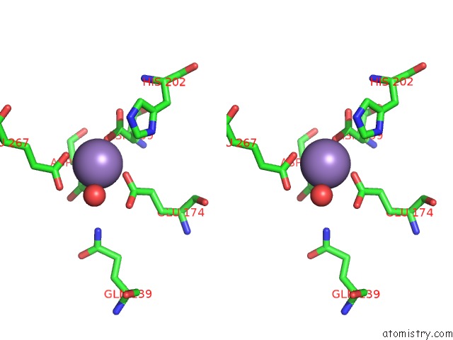
Stereo pair view
Mono view
Stereo pair view
A full contact list of Manganese with other atoms in the Mn binding
site number 2 of Crystal Structure of Sacuvde within 5.0Å range:
|
Reference:
E.M.Meulenbroek,
C.Peron Cane,
I.Jala,
S.Iwai,
G.F.Moolenaar,
N.Goosen,
N.S.Pannu.
Uv Damage Endonuclease Employs A Novel Dual-Dinucleotide Flipping Mechanism to Recognize Different Dna Lesions. Nucleic Acids Res. V. 41 1363 2013.
ISSN: ISSN 0305-1048
PubMed: 23221644
DOI: 10.1093/NAR/GKS1127
Page generated: Sat Oct 5 18:00:37 2024
ISSN: ISSN 0305-1048
PubMed: 23221644
DOI: 10.1093/NAR/GKS1127
Last articles
K in 7M2HK in 7MDJ
K in 7MJQ
K in 7MHR
K in 7MJO
K in 7M0D
K in 7M89
K in 7M83
K in 7M2I
K in 7M7T