Manganese »
PDB 3rla-3sx3 »
3sjt »
Manganese in PDB 3sjt: Crystal Structure of Human Arginase I in Complex with the Inhibitor Me-Abh, Resolution 1.60 A, Twinned Structure
Enzymatic activity of Crystal Structure of Human Arginase I in Complex with the Inhibitor Me-Abh, Resolution 1.60 A, Twinned Structure
All present enzymatic activity of Crystal Structure of Human Arginase I in Complex with the Inhibitor Me-Abh, Resolution 1.60 A, Twinned Structure:
3.5.3.1;
3.5.3.1;
Protein crystallography data
The structure of Crystal Structure of Human Arginase I in Complex with the Inhibitor Me-Abh, Resolution 1.60 A, Twinned Structure, PDB code: 3sjt
was solved by
L.Di Costanzo,
D.W.Christianson,
with X-Ray Crystallography technique. A brief refinement statistics is given in the table below:
| Resolution Low / High (Å) | 34.07 / 1.60 |
| Space group | P 3 |
| Cell size a, b, c (Å), α, β, γ (°) | 90.337, 90.337, 69.329, 90.00, 90.00, 120.00 |
| R / Rfree (%) | 12.9 / 16.3 |
Manganese Binding Sites:
The binding sites of Manganese atom in the Crystal Structure of Human Arginase I in Complex with the Inhibitor Me-Abh, Resolution 1.60 A, Twinned Structure
(pdb code 3sjt). This binding sites where shown within
5.0 Angstroms radius around Manganese atom.
In total 4 binding sites of Manganese where determined in the Crystal Structure of Human Arginase I in Complex with the Inhibitor Me-Abh, Resolution 1.60 A, Twinned Structure, PDB code: 3sjt:
Jump to Manganese binding site number: 1; 2; 3; 4;
In total 4 binding sites of Manganese where determined in the Crystal Structure of Human Arginase I in Complex with the Inhibitor Me-Abh, Resolution 1.60 A, Twinned Structure, PDB code: 3sjt:
Jump to Manganese binding site number: 1; 2; 3; 4;
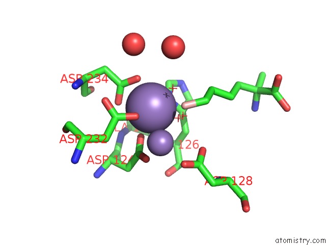
Manganese binding site 1 out of 4 in 3sjt
Go back to
Manganese binding site 1 out
of 4 in the Crystal Structure of Human Arginase I in Complex with the Inhibitor Me-Abh, Resolution 1.60 A, Twinned Structure
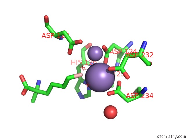
Mono view
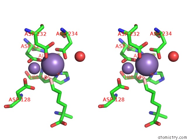
Stereo pair view
Mono view
Stereo pair view
A full contact list of Manganese with other atoms in the Mn binding
site number 1 of Crystal Structure of Human Arginase I in Complex with the Inhibitor Me-Abh, Resolution 1.60 A, Twinned Structure within 5.0Å range:
|
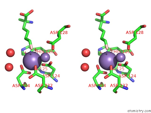
Manganese binding site 2 out of 4 in 3sjt
Go back to
Manganese binding site 2 out
of 4 in the Crystal Structure of Human Arginase I in Complex with the Inhibitor Me-Abh, Resolution 1.60 A, Twinned Structure
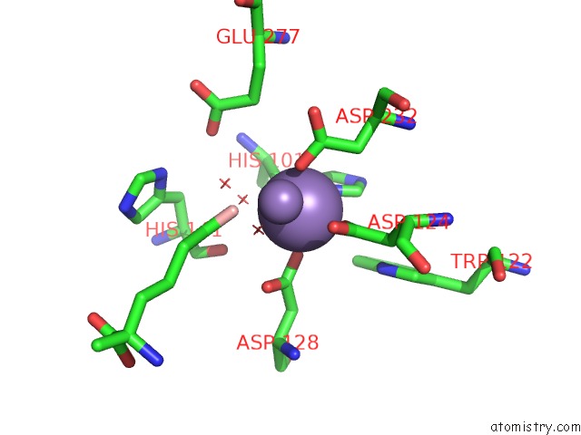
Mono view
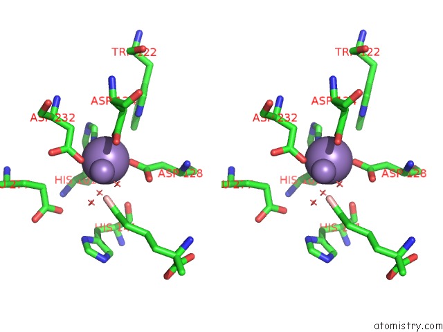
Stereo pair view
Mono view
Stereo pair view
A full contact list of Manganese with other atoms in the Mn binding
site number 2 of Crystal Structure of Human Arginase I in Complex with the Inhibitor Me-Abh, Resolution 1.60 A, Twinned Structure within 5.0Å range:
|
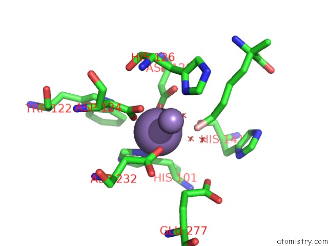
Manganese binding site 3 out of 4 in 3sjt
Go back to
Manganese binding site 3 out
of 4 in the Crystal Structure of Human Arginase I in Complex with the Inhibitor Me-Abh, Resolution 1.60 A, Twinned Structure
Mono view
Stereo pair view
Mono view
Stereo pair view
A full contact list of Manganese with other atoms in the Mn binding
site number 3 of Crystal Structure of Human Arginase I in Complex with the Inhibitor Me-Abh, Resolution 1.60 A, Twinned Structure within 5.0Å range:
|
Manganese binding site 4 out of 4 in 3sjt
Go back to
Manganese binding site 4 out
of 4 in the Crystal Structure of Human Arginase I in Complex with the Inhibitor Me-Abh, Resolution 1.60 A, Twinned Structure
Mono view
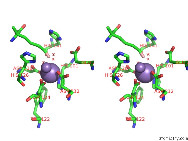
Stereo pair view
Mono view
Stereo pair view
A full contact list of Manganese with other atoms in the Mn binding
site number 4 of Crystal Structure of Human Arginase I in Complex with the Inhibitor Me-Abh, Resolution 1.60 A, Twinned Structure within 5.0Å range:
|
Reference:
M.Ilies,
L.Di Costanzo,
D.P.Dowling,
K.J.Thorn,
D.W.Christianson.
Binding of Alpha,Alpha-Disubstituted Amino Acids to Arginase Suggests New Avenues For Inhibitor Design J.Med.Chem. 2011.
ISSN: ISSN 0022-2623
PubMed: 21728378
DOI: 10.1021/JM200443B
Page generated: Sat Oct 5 17:53:23 2024
ISSN: ISSN 0022-2623
PubMed: 21728378
DOI: 10.1021/JM200443B
Last articles
Mg in 1JBZMg in 1JBW
Mg in 1JBV
Mg in 1JBK
Mg in 1JAX
Mg in 1JAH
Mg in 1J97
Mg in 1J9J
Mg in 1J8L
Mg in 1J7U