Manganese »
PDB 3q7c-3rl4 »
3qvn »
Manganese in PDB 3qvn: Crystal Structure of Cytosolic MNSOD3 From Candida Albicans
Protein crystallography data
The structure of Crystal Structure of Cytosolic MNSOD3 From Candida Albicans, PDB code: 3qvn
was solved by
Y.Sheng,
D.Cascio,
J.S.Valentine,
with X-Ray Crystallography technique. A brief refinement statistics is given in the table below:
| Resolution Low / High (Å) | 44.65 / 2.60 |
| Space group | P 64 2 2 |
| Cell size a, b, c (Å), α, β, γ (°) | 77.134, 77.134, 120.080, 90.00, 90.00, 120.00 |
| R / Rfree (%) | 21.2 / 27.3 |
Manganese Binding Sites:
The binding sites of Manganese atom in the Crystal Structure of Cytosolic MNSOD3 From Candida Albicans
(pdb code 3qvn). This binding sites where shown within
5.0 Angstroms radius around Manganese atom.
In total only one binding site of Manganese was determined in the Crystal Structure of Cytosolic MNSOD3 From Candida Albicans, PDB code: 3qvn:
In total only one binding site of Manganese was determined in the Crystal Structure of Cytosolic MNSOD3 From Candida Albicans, PDB code: 3qvn:
Manganese binding site 1 out of 1 in 3qvn
Go back to
Manganese binding site 1 out
of 1 in the Crystal Structure of Cytosolic MNSOD3 From Candida Albicans
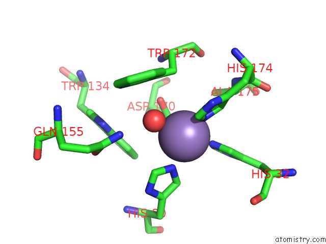
Mono view
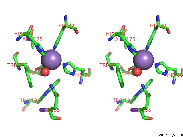
Stereo pair view
Mono view
Stereo pair view
A full contact list of Manganese with other atoms in the Mn binding
site number 1 of Crystal Structure of Cytosolic MNSOD3 From Candida Albicans within 5.0Å range:
|
Reference:
Y.Sheng,
T.A.Stich,
K.Barnese,
E.B.Gralla,
D.Cascio,
R.D.Britt,
D.E.Cabelli,
J.S.Valentine.
Comparison of Two Yeast Mnsods: Mitochondrial Saccharomyces Cerevisiae Versus Cytosolic Candida Albicans. J.Am.Chem.Soc. V. 133 20878 2011.
ISSN: ISSN 0002-7863
PubMed: 22077216
DOI: 10.1021/JA2077476
Page generated: Sat Oct 5 17:42:38 2024
ISSN: ISSN 0002-7863
PubMed: 22077216
DOI: 10.1021/JA2077476
Last articles
Mg in 3G10Mg in 3G0H
Mg in 3FXG
Mg in 3FYY
Mg in 3FZI
Mg in 3FYN
Mg in 3FYI
Mg in 3FYH
Mg in 3FYE
Mg in 3FXX