Manganese »
PDB 3q7c-3rl4 »
3qfo »
Manganese in PDB 3qfo: Crystal Structure of Streptococcal Asymmetric AP4A Hydrolase and Phosphodiesterase SPR1479/Saph Im Complex with Amp
Protein crystallography data
The structure of Crystal Structure of Streptococcal Asymmetric AP4A Hydrolase and Phosphodiesterase SPR1479/Saph Im Complex with Amp, PDB code: 3qfo
was solved by
Y.L.Jiang,
J.W.Zhang,
W.L.Yu,
W.Cheng,
C.C.Zhang,
C.Z.Zhou,
Y.Chen,
with X-Ray Crystallography technique. A brief refinement statistics is given in the table below:
| Resolution Low / High (Å) | 50.00 / 2.20 |
| Space group | P 21 21 21 |
| Cell size a, b, c (Å), α, β, γ (°) | 57.654, 74.940, 149.130, 90.00, 90.00, 90.00 |
| R / Rfree (%) | 18.5 / 24.5 |
Other elements in 3qfo:
The structure of Crystal Structure of Streptococcal Asymmetric AP4A Hydrolase and Phosphodiesterase SPR1479/Saph Im Complex with Amp also contains other interesting chemical elements:
| Iron | (Fe) | 2 atoms |
Manganese Binding Sites:
The binding sites of Manganese atom in the Crystal Structure of Streptococcal Asymmetric AP4A Hydrolase and Phosphodiesterase SPR1479/Saph Im Complex with Amp
(pdb code 3qfo). This binding sites where shown within
5.0 Angstroms radius around Manganese atom.
In total 2 binding sites of Manganese where determined in the Crystal Structure of Streptococcal Asymmetric AP4A Hydrolase and Phosphodiesterase SPR1479/Saph Im Complex with Amp, PDB code: 3qfo:
Jump to Manganese binding site number: 1; 2;
In total 2 binding sites of Manganese where determined in the Crystal Structure of Streptococcal Asymmetric AP4A Hydrolase and Phosphodiesterase SPR1479/Saph Im Complex with Amp, PDB code: 3qfo:
Jump to Manganese binding site number: 1; 2;
Manganese binding site 1 out of 2 in 3qfo
Go back to
Manganese binding site 1 out
of 2 in the Crystal Structure of Streptococcal Asymmetric AP4A Hydrolase and Phosphodiesterase SPR1479/Saph Im Complex with Amp
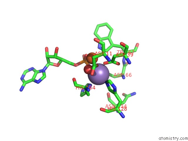
Mono view
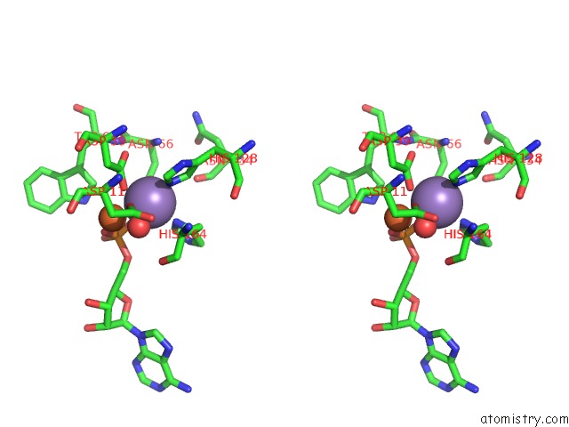
Stereo pair view
Mono view
Stereo pair view
A full contact list of Manganese with other atoms in the Mn binding
site number 1 of Crystal Structure of Streptococcal Asymmetric AP4A Hydrolase and Phosphodiesterase SPR1479/Saph Im Complex with Amp within 5.0Å range:
|
Manganese binding site 2 out of 2 in 3qfo
Go back to
Manganese binding site 2 out
of 2 in the Crystal Structure of Streptococcal Asymmetric AP4A Hydrolase and Phosphodiesterase SPR1479/Saph Im Complex with Amp
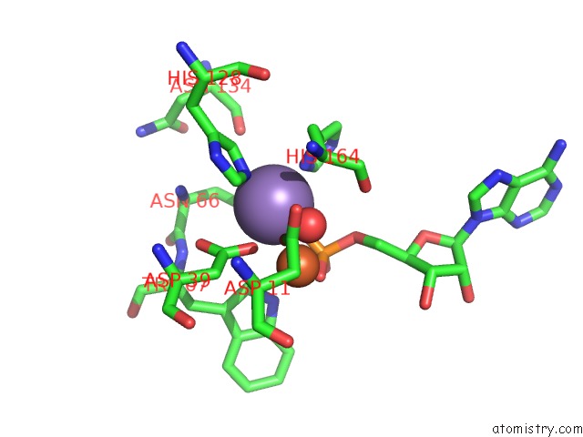
Mono view
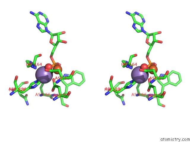
Stereo pair view
Mono view
Stereo pair view
A full contact list of Manganese with other atoms in the Mn binding
site number 2 of Crystal Structure of Streptococcal Asymmetric AP4A Hydrolase and Phosphodiesterase SPR1479/Saph Im Complex with Amp within 5.0Å range:
|
Reference:
Y.L.Jiang,
J.W.Zhang,
W.L.Yu,
W.Cheng,
C.C.Zhang,
C.Frolet,
A.-M.Di-Guilmi,
T.Vernet,
C.Z.Zhou,
Y.Chen.
Structural and Enzymatic Characterization of A Streptococcal Atp/Diadenosine Polyphosphate and Phosphodiester Hydrolase SPR1479/Saph To Be Published.
Page generated: Sat Oct 5 17:40:06 2024
Last articles
K in 3BGKK in 3BA6
K in 3B7I
K in 3B3C
K in 3B0N
K in 3B0H
K in 3B0M
K in 3B0L
K in 3B0G
K in 3B0J