Manganese »
PDB 3ot9-3q4q »
3q2v »
Manganese in PDB 3q2v: Crystal Structure of Mouse E-Cadherin Ectodomain
Protein crystallography data
The structure of Crystal Structure of Mouse E-Cadherin Ectodomain, PDB code: 3q2v
was solved by
X.Jin,
O.J.Harrison,
L.Shapiro,
with X-Ray Crystallography technique. A brief refinement statistics is given in the table below:
| Resolution Low / High (Å) | 19.92 / 3.40 |
| Space group | C 1 2 1 |
| Cell size a, b, c (Å), α, β, γ (°) | 119.140, 79.697, 176.001, 90.00, 98.56, 90.00 |
| R / Rfree (%) | 23 / 29.3 |
Other elements in 3q2v:
The structure of Crystal Structure of Mouse E-Cadherin Ectodomain also contains other interesting chemical elements:
| Calcium | (Ca) | 24 atoms |
Manganese Binding Sites:
The binding sites of Manganese atom in the Crystal Structure of Mouse E-Cadherin Ectodomain
(pdb code 3q2v). This binding sites where shown within
5.0 Angstroms radius around Manganese atom.
In total 2 binding sites of Manganese where determined in the Crystal Structure of Mouse E-Cadherin Ectodomain, PDB code: 3q2v:
Jump to Manganese binding site number: 1; 2;
In total 2 binding sites of Manganese where determined in the Crystal Structure of Mouse E-Cadherin Ectodomain, PDB code: 3q2v:
Jump to Manganese binding site number: 1; 2;
Manganese binding site 1 out of 2 in 3q2v
Go back to
Manganese binding site 1 out
of 2 in the Crystal Structure of Mouse E-Cadherin Ectodomain
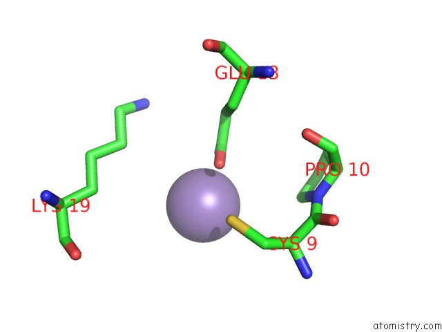
Mono view
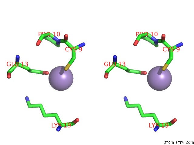
Stereo pair view
Mono view
Stereo pair view
A full contact list of Manganese with other atoms in the Mn binding
site number 1 of Crystal Structure of Mouse E-Cadherin Ectodomain within 5.0Å range:
|
Manganese binding site 2 out of 2 in 3q2v
Go back to
Manganese binding site 2 out
of 2 in the Crystal Structure of Mouse E-Cadherin Ectodomain
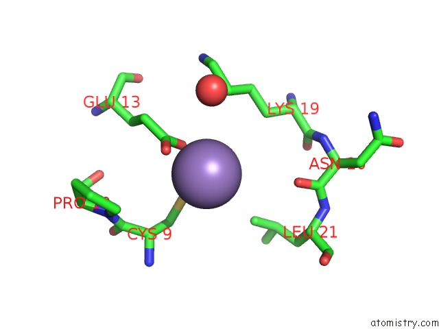
Mono view
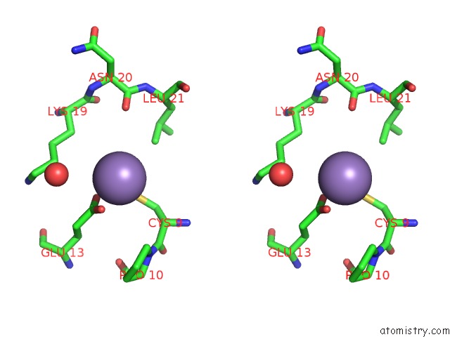
Stereo pair view
Mono view
Stereo pair view
A full contact list of Manganese with other atoms in the Mn binding
site number 2 of Crystal Structure of Mouse E-Cadherin Ectodomain within 5.0Å range:
|
Reference:
O.J.Harrison,
X.Jin,
S.Hong,
F.Bahna,
G.Ahlsen,
J.Brasch,
Y.Wu,
J.Vendome,
K.Felsovalyi,
C.M.Hampton,
R.B.Troyanovsky,
A.Ben-Shaul,
J.Frank,
S.M.Troyanovsky,
L.Shapiro,
B.Honig.
The Extracellular Architecture of Adherens Junctions Revealed By Crystal Structures of Type I Cadherins. Structure V. 19 244 2011.
ISSN: ISSN 0969-2126
PubMed: 21300292
DOI: 10.1016/J.STR.2010.11.016
Page generated: Sat Oct 5 17:35:38 2024
ISSN: ISSN 0969-2126
PubMed: 21300292
DOI: 10.1016/J.STR.2010.11.016
Last articles
K in 8OLWK in 8OLJ
K in 8OFD
K in 8OEO
K in 8OED
K in 8OEH
K in 8K1Z
K in 8K1U
K in 8K1V
K in 8K7W