Manganese »
PDB 3ot9-3q4q »
3pzt »
Manganese in PDB 3pzt: Structure of the Endo-1,4-Beta-Glucanase From Bacillus Subtilis 168 with Manganese(II) Ion
Enzymatic activity of Structure of the Endo-1,4-Beta-Glucanase From Bacillus Subtilis 168 with Manganese(II) Ion
All present enzymatic activity of Structure of the Endo-1,4-Beta-Glucanase From Bacillus Subtilis 168 with Manganese(II) Ion:
3.2.1.4;
3.2.1.4;
Protein crystallography data
The structure of Structure of the Endo-1,4-Beta-Glucanase From Bacillus Subtilis 168 with Manganese(II) Ion, PDB code: 3pzt
was solved by
C.R.Santos,
J.H.Paiva,
P.K.Akao,
A.N.Meza,
J.C.Silva,
F.M.Squina,
R.J.Ward,
R.Ruller,
M.T.Murakami,
with X-Ray Crystallography technique. A brief refinement statistics is given in the table below:
| Resolution Low / High (Å) | 42.40 / 1.97 |
| Space group | P 21 21 21 |
| Cell size a, b, c (Å), α, β, γ (°) | 49.526, 110.617, 122.262, 90.00, 90.00, 90.00 |
| R / Rfree (%) | 16.4 / 20.6 |
Manganese Binding Sites:
The binding sites of Manganese atom in the Structure of the Endo-1,4-Beta-Glucanase From Bacillus Subtilis 168 with Manganese(II) Ion
(pdb code 3pzt). This binding sites where shown within
5.0 Angstroms radius around Manganese atom.
In total 2 binding sites of Manganese where determined in the Structure of the Endo-1,4-Beta-Glucanase From Bacillus Subtilis 168 with Manganese(II) Ion, PDB code: 3pzt:
Jump to Manganese binding site number: 1; 2;
In total 2 binding sites of Manganese where determined in the Structure of the Endo-1,4-Beta-Glucanase From Bacillus Subtilis 168 with Manganese(II) Ion, PDB code: 3pzt:
Jump to Manganese binding site number: 1; 2;
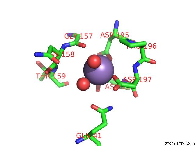
Manganese binding site 1 out of 2 in 3pzt
Go back to
Manganese binding site 1 out
of 2 in the Structure of the Endo-1,4-Beta-Glucanase From Bacillus Subtilis 168 with Manganese(II) Ion
Mono view
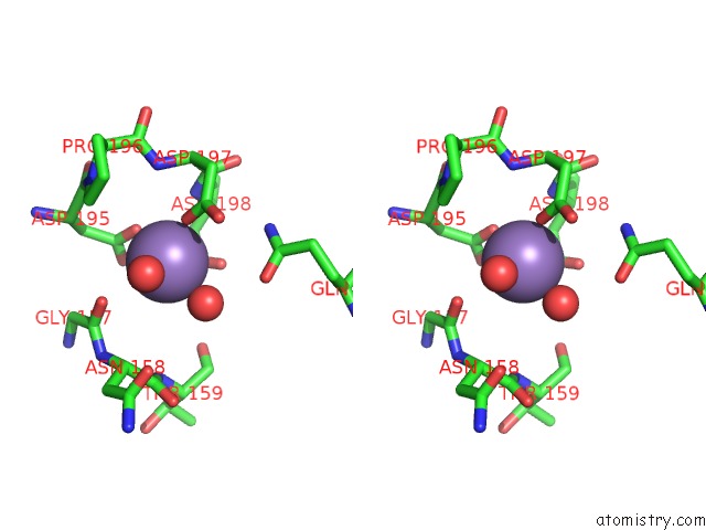
Stereo pair view
Mono view
Stereo pair view
A full contact list of Manganese with other atoms in the Mn binding
site number 1 of Structure of the Endo-1,4-Beta-Glucanase From Bacillus Subtilis 168 with Manganese(II) Ion within 5.0Å range:
|
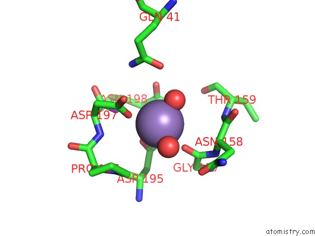
Manganese binding site 2 out of 2 in 3pzt
Go back to
Manganese binding site 2 out
of 2 in the Structure of the Endo-1,4-Beta-Glucanase From Bacillus Subtilis 168 with Manganese(II) Ion
Mono view
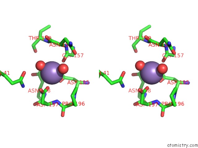
Stereo pair view
Mono view
Stereo pair view
A full contact list of Manganese with other atoms in the Mn binding
site number 2 of Structure of the Endo-1,4-Beta-Glucanase From Bacillus Subtilis 168 with Manganese(II) Ion within 5.0Å range:
|
Reference:
C.R.Santos,
J.H.Paiva,
M.L.Sforca,
J.L.Neves,
R.Z.Navarro,
J.Cota,
P.K.Akao,
Z.B.Hoffmam,
A.N.Meza,
J.H.Smetana,
M.L.Nogueira,
I.Polikarpov,
J.Xavier-Neto,
F.M.Squina,
R.J.Ward,
R.Ruller,
A.C.Zeri,
M.T.Murakami.
Dissecting Structure-Function-Stability Relationships of A Thermostable GH5-CBM3 Cellulase From Bacillus Subtilis 168. Biochem.J. V. 441 95 2012.
ISSN: ISSN 0264-6021
PubMed: 21880019
DOI: 10.1042/BJ20110869
Page generated: Sat Oct 5 17:35:24 2024
ISSN: ISSN 0264-6021
PubMed: 21880019
DOI: 10.1042/BJ20110869
Last articles
K in 4TODK in 4TOA
K in 4TO9
K in 4TM0
K in 4TMZ
K in 4TLZ
K in 4TLX
K in 4RVO
K in 4RVN
K in 4TKX