Manganese »
PDB 3ot9-3q4q »
3oul »
Manganese in PDB 3oul: Crystal Structure of Toxoflavin-Degrading Enzyme in A Substrate-Free Form
Protein crystallography data
The structure of Crystal Structure of Toxoflavin-Degrading Enzyme in A Substrate-Free Form, PDB code: 3oul
was solved by
M.I.Kim,
S.Rhee,
with X-Ray Crystallography technique. A brief refinement statistics is given in the table below:
| Resolution Low / High (Å) | 50.00 / 1.60 |
| Space group | H 3 |
| Cell size a, b, c (Å), α, β, γ (°) | 110.377, 110.377, 56.787, 90.00, 90.00, 120.00 |
| R / Rfree (%) | 19.5 / 22 |
Manganese Binding Sites:
The binding sites of Manganese atom in the Crystal Structure of Toxoflavin-Degrading Enzyme in A Substrate-Free Form
(pdb code 3oul). This binding sites where shown within
5.0 Angstroms radius around Manganese atom.
In total only one binding site of Manganese was determined in the Crystal Structure of Toxoflavin-Degrading Enzyme in A Substrate-Free Form, PDB code: 3oul:
In total only one binding site of Manganese was determined in the Crystal Structure of Toxoflavin-Degrading Enzyme in A Substrate-Free Form, PDB code: 3oul:
Manganese binding site 1 out of 1 in 3oul
Go back to
Manganese binding site 1 out
of 1 in the Crystal Structure of Toxoflavin-Degrading Enzyme in A Substrate-Free Form
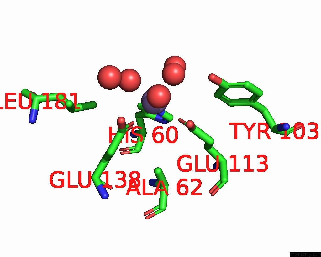
Mono view
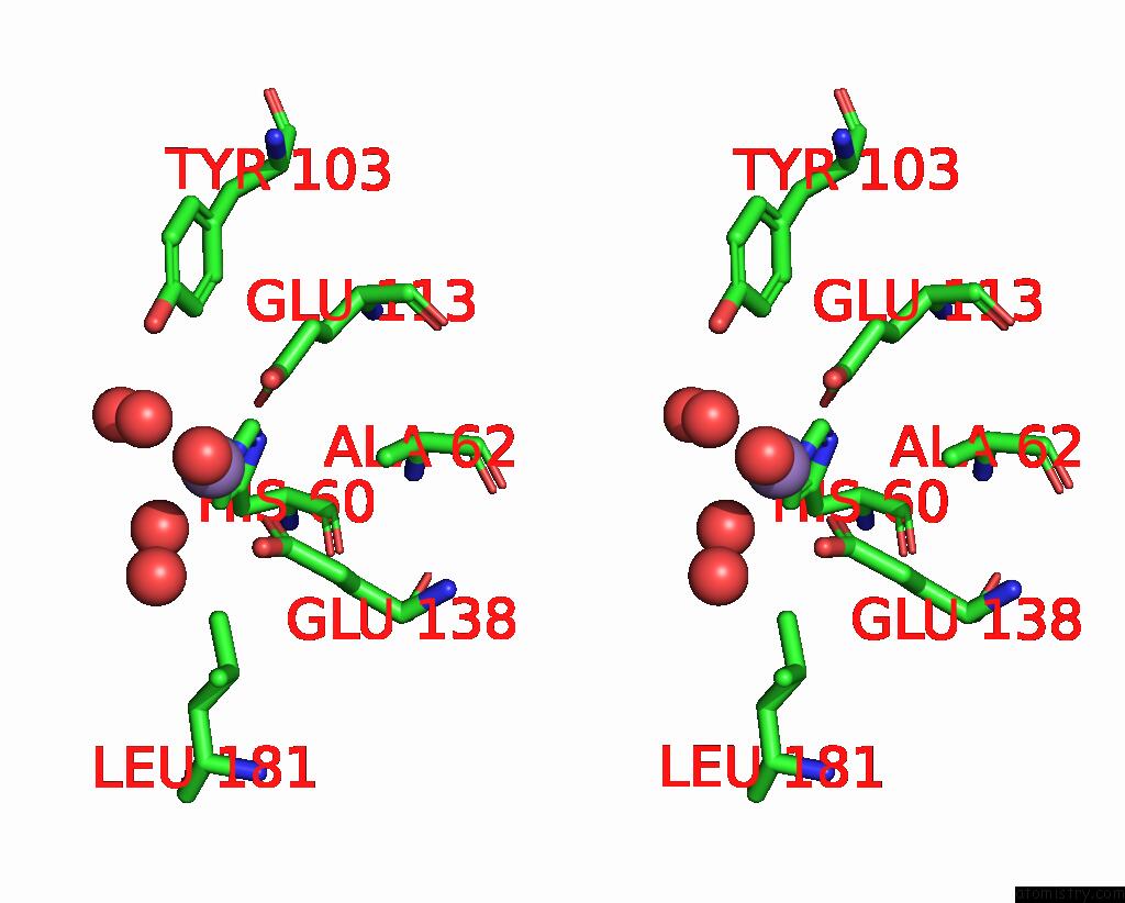
Stereo pair view
Mono view
Stereo pair view
A full contact list of Manganese with other atoms in the Mn binding
site number 1 of Crystal Structure of Toxoflavin-Degrading Enzyme in A Substrate-Free Form within 5.0Å range:
|
Reference:
W.S.Jung,
J.Lee,
M.I.Kim,
J.Ma,
T.Nagamatsu,
E.Goo,
H.Kim,
I.Hwang,
J.Han,
S.Rhee.
Structural and Functional Analysis of Phytotoxin Toxoflavin-Degrading Enzyme Plos One V. 6 22443 2011.
ISSN: ESSN 1932-6203
PubMed: 21799856
DOI: 10.1371/JOURNAL.PONE.0022443
Page generated: Sat Oct 5 17:27:56 2024
ISSN: ESSN 1932-6203
PubMed: 21799856
DOI: 10.1371/JOURNAL.PONE.0022443
Last articles
K in 4U75K in 4U73
K in 4U71
K in 4U70
K in 4U6Z
K in 4TS0
K in 4U6W
K in 4U6J
K in 4U5M
K in 4U6C