Manganese »
PDB 3n39-3orm »
3orl »
Manganese in PDB 3orl: Mycobacterium Tuberculosis Pknb Kinase Domain L33D Mutant (Crystal Form 3)
Protein crystallography data
The structure of Mycobacterium Tuberculosis Pknb Kinase Domain L33D Mutant (Crystal Form 3), PDB code: 3orl
was solved by
N.Echols,
T.N.Lombana,
N.D.Thomsen,
H.-L.Ng,
T.Alber,
Tb Structuralgenomics Consortium (Tbsgc),
with X-Ray Crystallography technique. A brief refinement statistics is given in the table below:
| Resolution Low / High (Å) | 20.00 / 2.90 |
| Space group | P 21 21 21 |
| Cell size a, b, c (Å), α, β, γ (°) | 40.191, 50.633, 132.762, 90.00, 90.00, 90.00 |
| R / Rfree (%) | 21.1 / 27.7 |
Manganese Binding Sites:
The binding sites of Manganese atom in the Mycobacterium Tuberculosis Pknb Kinase Domain L33D Mutant (Crystal Form 3)
(pdb code 3orl). This binding sites where shown within
5.0 Angstroms radius around Manganese atom.
In total only one binding site of Manganese was determined in the Mycobacterium Tuberculosis Pknb Kinase Domain L33D Mutant (Crystal Form 3), PDB code: 3orl:
In total only one binding site of Manganese was determined in the Mycobacterium Tuberculosis Pknb Kinase Domain L33D Mutant (Crystal Form 3), PDB code: 3orl:
Manganese binding site 1 out of 1 in 3orl
Go back to
Manganese binding site 1 out
of 1 in the Mycobacterium Tuberculosis Pknb Kinase Domain L33D Mutant (Crystal Form 3)
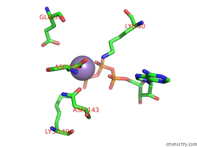
Mono view
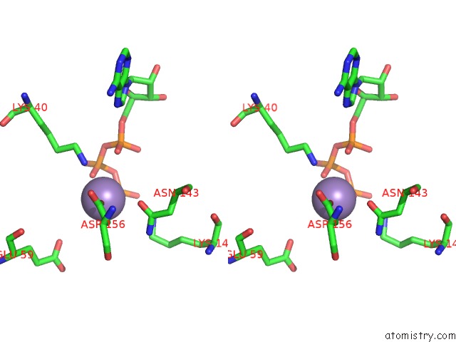
Stereo pair view
Mono view
Stereo pair view
A full contact list of Manganese with other atoms in the Mn binding
site number 1 of Mycobacterium Tuberculosis Pknb Kinase Domain L33D Mutant (Crystal Form 3) within 5.0Å range:
|
Reference:
T.N.Lombana,
N.Echols,
M.C.Good,
N.D.Thomsen,
H.-L.Ng,
A.E.Greenstein,
A.M.Falick,
D.S.King,
T.Alber.
Allosteric Activation Mechanism of the Mycobacterium Tuberculosis Receptor Ser/Thr Kinase, Pknb Structure V. 18 1667 2010.
ISSN: ISSN 0969-2126
Page generated: Sat Oct 5 17:26:37 2024
ISSN: ISSN 0969-2126
Last articles
Kr in 1C6MKr in 1QTK
Kr in 1C6J
Kr in 1C6Q
Kr in 1C6D
Kr in 1C6G
Kr in 1C67
Kr in 1C61
Kr in 1C6A
Kr in 1C64