Manganese »
PDB 3n39-3orm »
3nvt »
Manganese in PDB 3nvt: 1.95 Angstrom Crystal Structure of A Bifunctional 3-Deoxy-7- Phosphoheptulonate Synthase/Chorismate Mutase (Aroa) From Listeria Monocytogenes Egd-E
Enzymatic activity of 1.95 Angstrom Crystal Structure of A Bifunctional 3-Deoxy-7- Phosphoheptulonate Synthase/Chorismate Mutase (Aroa) From Listeria Monocytogenes Egd-E
All present enzymatic activity of 1.95 Angstrom Crystal Structure of A Bifunctional 3-Deoxy-7- Phosphoheptulonate Synthase/Chorismate Mutase (Aroa) From Listeria Monocytogenes Egd-E:
2.5.1.54; 5.4.99.5;
2.5.1.54; 5.4.99.5;
Protein crystallography data
The structure of 1.95 Angstrom Crystal Structure of A Bifunctional 3-Deoxy-7- Phosphoheptulonate Synthase/Chorismate Mutase (Aroa) From Listeria Monocytogenes Egd-E, PDB code: 3nvt
was solved by
A.S.Halavaty,
S.H.Light,
G.Minasov,
L.Shuvalova,
K.Kwon,
W.F.Anderson,
Center For Structural Genomics Of Infectious Diseases (Csgid),
with X-Ray Crystallography technique. A brief refinement statistics is given in the table below:
| Resolution Low / High (Å) | 29.04 / 1.95 |
| Space group | C 1 2 1 |
| Cell size a, b, c (Å), α, β, γ (°) | 111.570, 111.787, 81.049, 90.00, 127.63, 90.00 |
| R / Rfree (%) | 15.4 / 19.8 |
Manganese Binding Sites:
The binding sites of Manganese atom in the 1.95 Angstrom Crystal Structure of A Bifunctional 3-Deoxy-7- Phosphoheptulonate Synthase/Chorismate Mutase (Aroa) From Listeria Monocytogenes Egd-E
(pdb code 3nvt). This binding sites where shown within
5.0 Angstroms radius around Manganese atom.
In total 4 binding sites of Manganese where determined in the 1.95 Angstrom Crystal Structure of A Bifunctional 3-Deoxy-7- Phosphoheptulonate Synthase/Chorismate Mutase (Aroa) From Listeria Monocytogenes Egd-E, PDB code: 3nvt:
Jump to Manganese binding site number: 1; 2; 3; 4;
In total 4 binding sites of Manganese where determined in the 1.95 Angstrom Crystal Structure of A Bifunctional 3-Deoxy-7- Phosphoheptulonate Synthase/Chorismate Mutase (Aroa) From Listeria Monocytogenes Egd-E, PDB code: 3nvt:
Jump to Manganese binding site number: 1; 2; 3; 4;
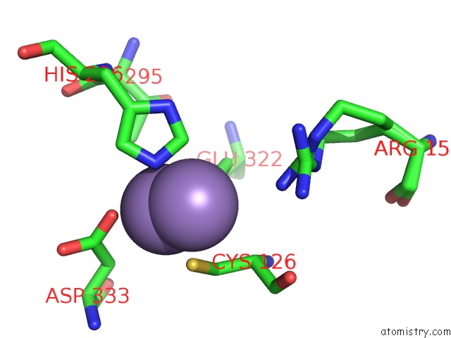
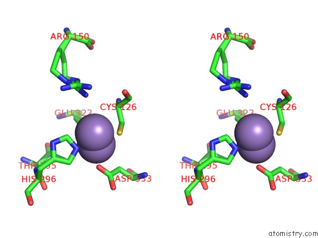
Manganese binding site 1 out of 4 in 3nvt
Go back to
Manganese binding site 1 out
of 4 in the 1.95 Angstrom Crystal Structure of A Bifunctional 3-Deoxy-7- Phosphoheptulonate Synthase/Chorismate Mutase (Aroa) From Listeria Monocytogenes Egd-E
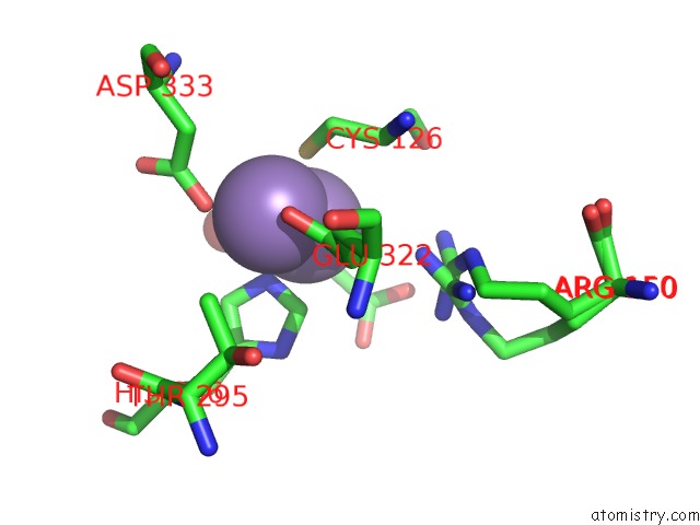
Mono view
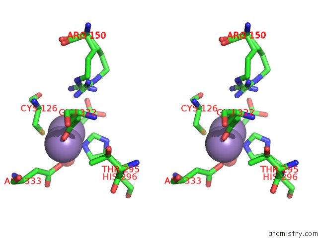
Stereo pair view
Mono view
Stereo pair view
A full contact list of Manganese with other atoms in the Mn binding
site number 1 of 1.95 Angstrom Crystal Structure of A Bifunctional 3-Deoxy-7- Phosphoheptulonate Synthase/Chorismate Mutase (Aroa) From Listeria Monocytogenes Egd-E within 5.0Å range:
|
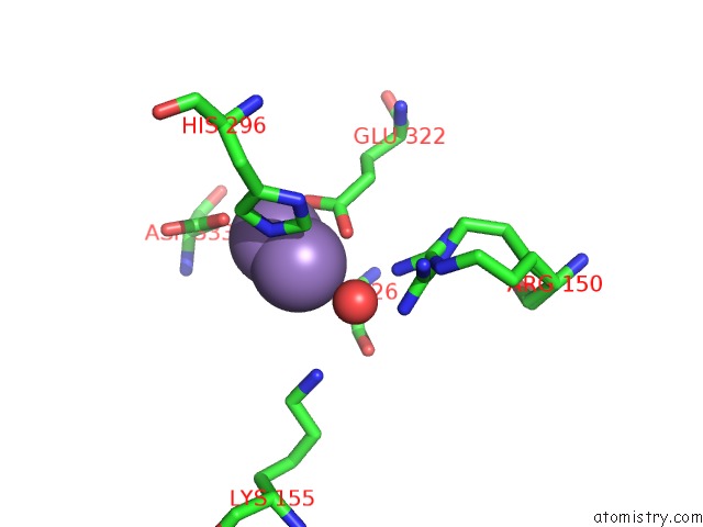
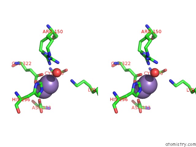
Manganese binding site 2 out of 4 in 3nvt
Go back to
Manganese binding site 2 out
of 4 in the 1.95 Angstrom Crystal Structure of A Bifunctional 3-Deoxy-7- Phosphoheptulonate Synthase/Chorismate Mutase (Aroa) From Listeria Monocytogenes Egd-E
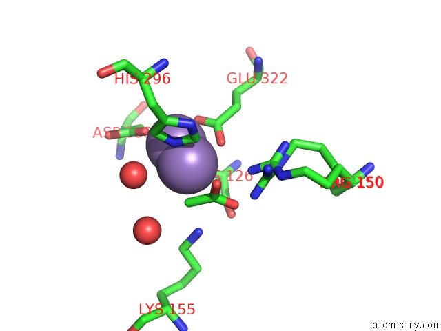
Mono view
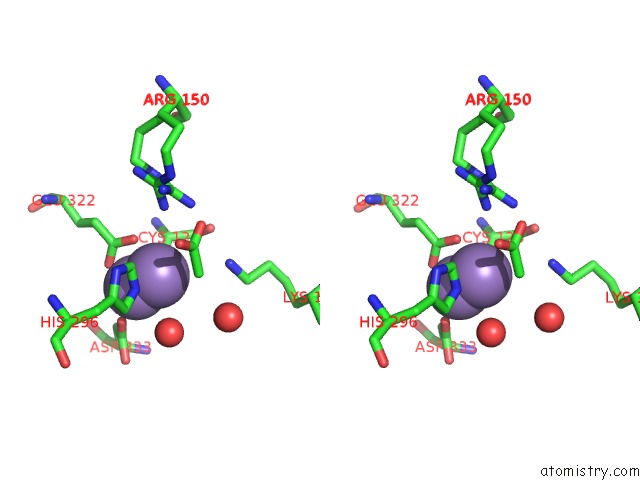
Stereo pair view
Mono view
Stereo pair view
A full contact list of Manganese with other atoms in the Mn binding
site number 2 of 1.95 Angstrom Crystal Structure of A Bifunctional 3-Deoxy-7- Phosphoheptulonate Synthase/Chorismate Mutase (Aroa) From Listeria Monocytogenes Egd-E within 5.0Å range:
|
Manganese binding site 3 out of 4 in 3nvt
Go back to
Manganese binding site 3 out
of 4 in the 1.95 Angstrom Crystal Structure of A Bifunctional 3-Deoxy-7- Phosphoheptulonate Synthase/Chorismate Mutase (Aroa) From Listeria Monocytogenes Egd-E
Mono view
Stereo pair view
Mono view
Stereo pair view
A full contact list of Manganese with other atoms in the Mn binding
site number 3 of 1.95 Angstrom Crystal Structure of A Bifunctional 3-Deoxy-7- Phosphoheptulonate Synthase/Chorismate Mutase (Aroa) From Listeria Monocytogenes Egd-E within 5.0Å range:
|
Manganese binding site 4 out of 4 in 3nvt
Go back to
Manganese binding site 4 out
of 4 in the 1.95 Angstrom Crystal Structure of A Bifunctional 3-Deoxy-7- Phosphoheptulonate Synthase/Chorismate Mutase (Aroa) From Listeria Monocytogenes Egd-E
Mono view
Stereo pair view
Mono view
Stereo pair view
A full contact list of Manganese with other atoms in the Mn binding
site number 4 of 1.95 Angstrom Crystal Structure of A Bifunctional 3-Deoxy-7- Phosphoheptulonate Synthase/Chorismate Mutase (Aroa) From Listeria Monocytogenes Egd-E within 5.0Å range:
|
Reference:
S.H.Light,
A.S.Halavaty,
G.Minasov,
L.Shuvalova,
W.F.Anderson.
Structural Analysis of A 3-Deoxy-D-Arabino-Heptulosonate 7-Phosphate Synthase with An N-Terminal Chorismate Mutase-Like Regulatory Domain. Protein Sci. V. 21 887 2012.
ISSN: ISSN 0961-8368
PubMed: 22505283
DOI: 10.1002/PRO.2075
Page generated: Sat Oct 5 17:21:27 2024
ISSN: ISSN 0961-8368
PubMed: 22505283
DOI: 10.1002/PRO.2075
Last articles
K in 8OLWK in 8OLJ
K in 8OFD
K in 8OEO
K in 8OED
K in 8OEH
K in 8K1Z
K in 8K1U
K in 8K1V
K in 8K7W