Manganese »
PDB 3n39-3orm »
3nio »
Manganese in PDB 3nio: Crystal Structure of Pseudomonas Aeruginosa Guanidinobutyrase
Enzymatic activity of Crystal Structure of Pseudomonas Aeruginosa Guanidinobutyrase
All present enzymatic activity of Crystal Structure of Pseudomonas Aeruginosa Guanidinobutyrase:
3.5.3.7;
3.5.3.7;
Protein crystallography data
The structure of Crystal Structure of Pseudomonas Aeruginosa Guanidinobutyrase, PDB code: 3nio
was solved by
S.J.Lee,
H.S.Kim,
D.J.Kim,
H.J.Yoon,
K.H.Kim,
J.Y.Yoon,
J.Y.Jang,
H.Im,
D.An,
S.W.Suh,
with X-Ray Crystallography technique. A brief refinement statistics is given in the table below:
| Resolution Low / High (Å) | 20.00 / 2.00 |
| Space group | P 1 21 1 |
| Cell size a, b, c (Å), α, β, γ (°) | 82.047, 158.954, 84.547, 90.00, 114.84, 90.00 |
| R / Rfree (%) | 23.3 / 25.1 |
Manganese Binding Sites:
Pages:
>>> Page 1 <<< Page 2, Binding sites: 11 - 12;Binding sites:
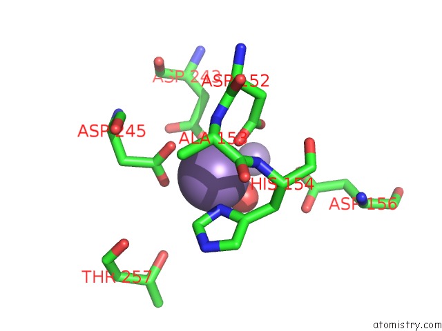
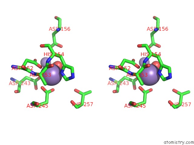
The binding sites of Manganese atom in the Crystal Structure of Pseudomonas Aeruginosa Guanidinobutyrase (pdb code 3nio). This binding sites where shown within 5.0 Angstroms radius around Manganese atom.In total 12 binding sites of Manganese where determined in the Crystal Structure of Pseudomonas Aeruginosa Guanidinobutyrase, PDB code: 3nio:
Jump to Manganese binding site number: 1; 2; 3; 4; 5; 6; 7; 8; 9; 10;
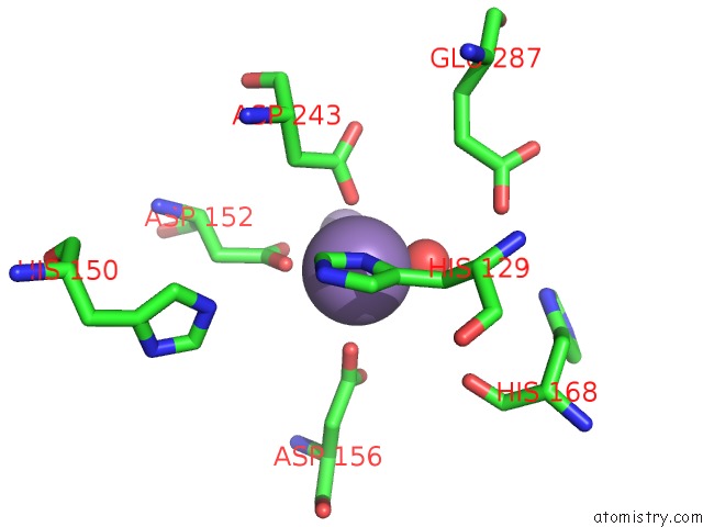
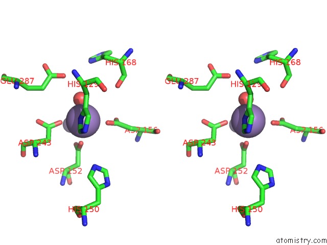
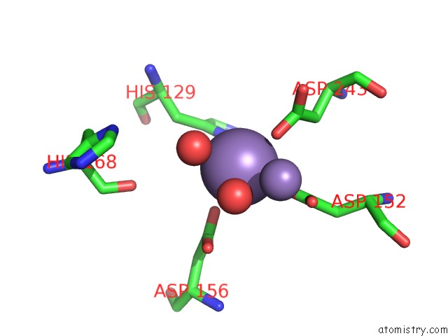
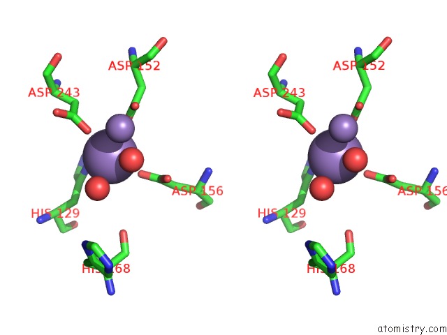
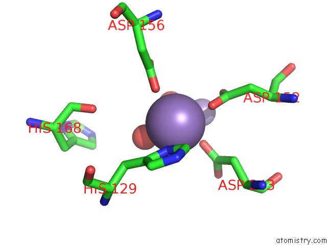
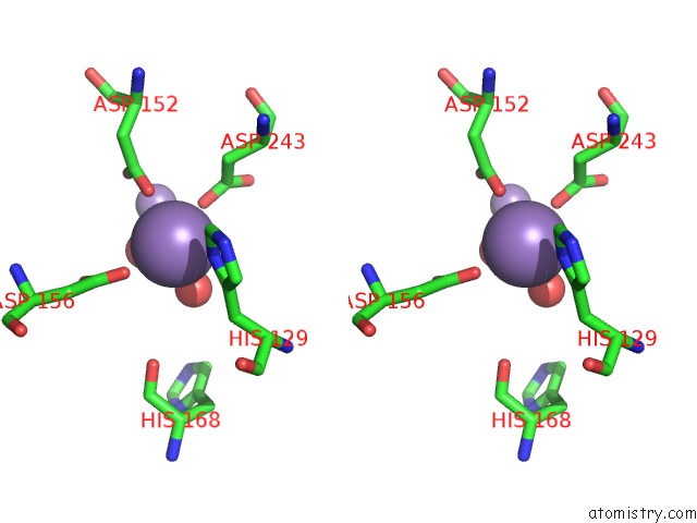
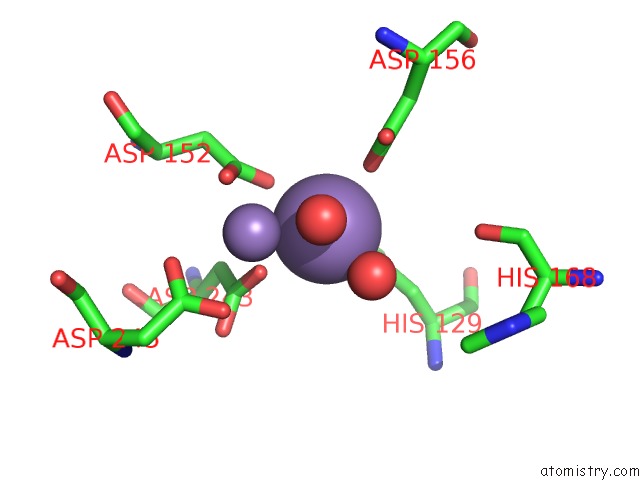
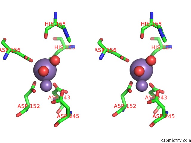
Manganese binding site 1 out of 12 in 3nio
Go back to
Manganese binding site 1 out
of 12 in the Crystal Structure of Pseudomonas Aeruginosa Guanidinobutyrase
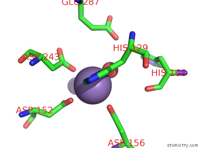
Mono view
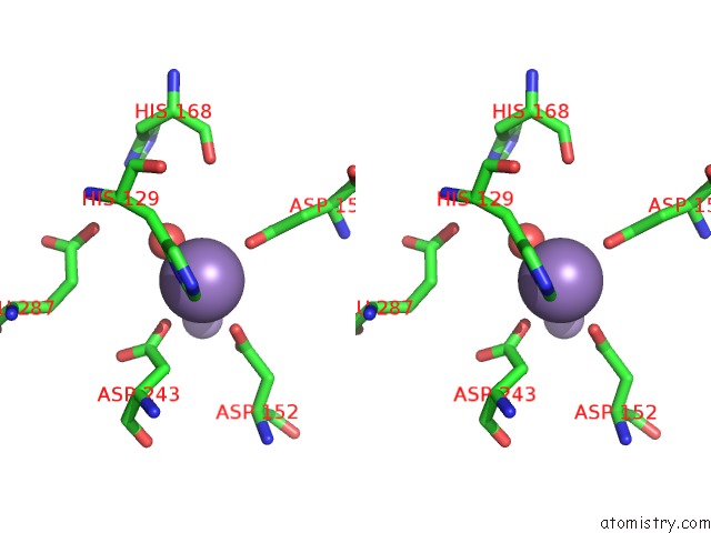
Stereo pair view
Mono view
Stereo pair view
A full contact list of Manganese with other atoms in the Mn binding
site number 1 of Crystal Structure of Pseudomonas Aeruginosa Guanidinobutyrase within 5.0Å range:
|
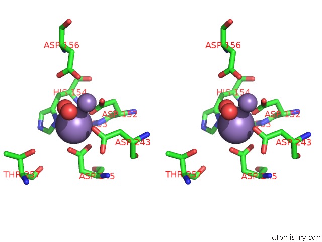
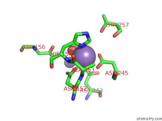
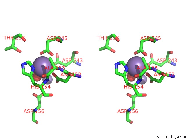
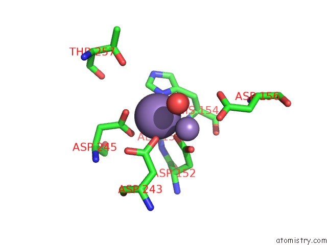
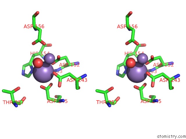
Manganese binding site 2 out of 12 in 3nio
Go back to
Manganese binding site 2 out
of 12 in the Crystal Structure of Pseudomonas Aeruginosa Guanidinobutyrase
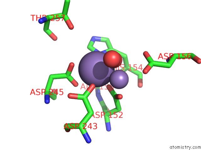
Mono view
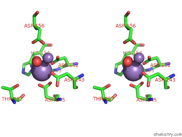
Stereo pair view
Mono view
Stereo pair view
A full contact list of Manganese with other atoms in the Mn binding
site number 2 of Crystal Structure of Pseudomonas Aeruginosa Guanidinobutyrase within 5.0Å range:
|
Manganese binding site 3 out of 12 in 3nio
Go back to
Manganese binding site 3 out
of 12 in the Crystal Structure of Pseudomonas Aeruginosa Guanidinobutyrase
Mono view
Stereo pair view
Mono view
Stereo pair view
A full contact list of Manganese with other atoms in the Mn binding
site number 3 of Crystal Structure of Pseudomonas Aeruginosa Guanidinobutyrase within 5.0Å range:
|
Manganese binding site 4 out of 12 in 3nio
Go back to
Manganese binding site 4 out
of 12 in the Crystal Structure of Pseudomonas Aeruginosa Guanidinobutyrase

Mono view
Stereo pair view
Mono view
Stereo pair view
A full contact list of Manganese with other atoms in the Mn binding
site number 4 of Crystal Structure of Pseudomonas Aeruginosa Guanidinobutyrase within 5.0Å range:
|
Manganese binding site 5 out of 12 in 3nio
Go back to
Manganese binding site 5 out
of 12 in the Crystal Structure of Pseudomonas Aeruginosa Guanidinobutyrase
Mono view
Stereo pair view
Mono view
Stereo pair view
A full contact list of Manganese with other atoms in the Mn binding
site number 5 of Crystal Structure of Pseudomonas Aeruginosa Guanidinobutyrase within 5.0Å range:
|
Manganese binding site 6 out of 12 in 3nio
Go back to
Manganese binding site 6 out
of 12 in the Crystal Structure of Pseudomonas Aeruginosa Guanidinobutyrase
Mono view
Stereo pair view
Mono view
Stereo pair view
A full contact list of Manganese with other atoms in the Mn binding
site number 6 of Crystal Structure of Pseudomonas Aeruginosa Guanidinobutyrase within 5.0Å range:
|
Manganese binding site 7 out of 12 in 3nio
Go back to
Manganese binding site 7 out
of 12 in the Crystal Structure of Pseudomonas Aeruginosa Guanidinobutyrase
Mono view
Stereo pair view
Mono view
Stereo pair view
A full contact list of Manganese with other atoms in the Mn binding
site number 7 of Crystal Structure of Pseudomonas Aeruginosa Guanidinobutyrase within 5.0Å range:
|
Manganese binding site 8 out of 12 in 3nio
Go back to
Manganese binding site 8 out
of 12 in the Crystal Structure of Pseudomonas Aeruginosa Guanidinobutyrase
Mono view
Stereo pair view
Mono view
Stereo pair view
A full contact list of Manganese with other atoms in the Mn binding
site number 8 of Crystal Structure of Pseudomonas Aeruginosa Guanidinobutyrase within 5.0Å range:
|
Manganese binding site 9 out of 12 in 3nio
Go back to
Manganese binding site 9 out
of 12 in the Crystal Structure of Pseudomonas Aeruginosa Guanidinobutyrase
Mono view
Stereo pair view
Mono view
Stereo pair view
A full contact list of Manganese with other atoms in the Mn binding
site number 9 of Crystal Structure of Pseudomonas Aeruginosa Guanidinobutyrase within 5.0Å range:
|
Manganese binding site 10 out of 12 in 3nio
Go back to
Manganese binding site 10 out
of 12 in the Crystal Structure of Pseudomonas Aeruginosa Guanidinobutyrase
Mono view
Stereo pair view
Mono view
Stereo pair view
A full contact list of Manganese with other atoms in the Mn binding
site number 10 of Crystal Structure of Pseudomonas Aeruginosa Guanidinobutyrase within 5.0Å range:
|
Reference:
S.J.Lee,
D.J.Kim,
H.S.Kim,
B.I.Lee,
H.J.Yoon,
J.Y.Yoon,
K.H.Kim,
J.Y.Jang,
H.N.Im,
D.R.An,
J.S.Song,
H.J.Kim,
S.W.Suh.
Crystal Structures of Pseudomonas Aeruginosa Guanidinobutyrase and Guanidinopropionase, Members of the Ureohydrolase Superfamily J.Struct.Biol. V. 175 329 2011.
ISSN: ISSN 1047-8477
PubMed: 21600989
DOI: 10.1016/J.JSB.2011.05.002
Page generated: Sat Oct 5 17:19:53 2024
ISSN: ISSN 1047-8477
PubMed: 21600989
DOI: 10.1016/J.JSB.2011.05.002
Last articles
Kr in 1C6MKr in 1QTK
Kr in 1C6J
Kr in 1C6Q
Kr in 1C6D
Kr in 1C6G
Kr in 1C67
Kr in 1C61
Kr in 1C6A
Kr in 1C64