Manganese »
PDB 3m0m-3n37 »
3mfw »
Manganese in PDB 3mfw: Crystal Structure of Human Arginase I in Complex with L-2- Aminohistidine and Sulphate
Enzymatic activity of Crystal Structure of Human Arginase I in Complex with L-2- Aminohistidine and Sulphate
All present enzymatic activity of Crystal Structure of Human Arginase I in Complex with L-2- Aminohistidine and Sulphate:
3.5.3.1;
3.5.3.1;
Protein crystallography data
The structure of Crystal Structure of Human Arginase I in Complex with L-2- Aminohistidine and Sulphate, PDB code: 3mfw
was solved by
L.Di Costanzo,
D.W.Christianson,
with X-Ray Crystallography technique. A brief refinement statistics is given in the table below:
| Resolution Low / High (Å) | 50.00 / 1.47 |
| Space group | P 3 |
| Cell size a, b, c (Å), α, β, γ (°) | 90.338, 90.338, 69.429, 90.00, 90.00, 120.00 |
| R / Rfree (%) | 14.9 / 16.2 |
Manganese Binding Sites:
The binding sites of Manganese atom in the Crystal Structure of Human Arginase I in Complex with L-2- Aminohistidine and Sulphate
(pdb code 3mfw). This binding sites where shown within
5.0 Angstroms radius around Manganese atom.
In total 4 binding sites of Manganese where determined in the Crystal Structure of Human Arginase I in Complex with L-2- Aminohistidine and Sulphate, PDB code: 3mfw:
Jump to Manganese binding site number: 1; 2; 3; 4;
In total 4 binding sites of Manganese where determined in the Crystal Structure of Human Arginase I in Complex with L-2- Aminohistidine and Sulphate, PDB code: 3mfw:
Jump to Manganese binding site number: 1; 2; 3; 4;
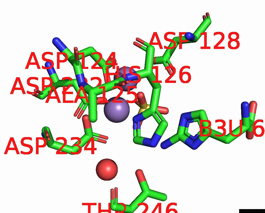
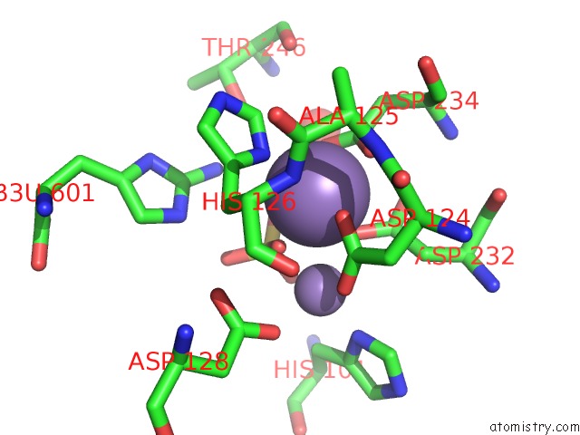
Manganese binding site 1 out of 4 in 3mfw
Go back to
Manganese binding site 1 out
of 4 in the Crystal Structure of Human Arginase I in Complex with L-2- Aminohistidine and Sulphate
Mono view
Stereo pair view
Mono view
Stereo pair view
A full contact list of Manganese with other atoms in the Mn binding
site number 1 of Crystal Structure of Human Arginase I in Complex with L-2- Aminohistidine and Sulphate within 5.0Å range:
|
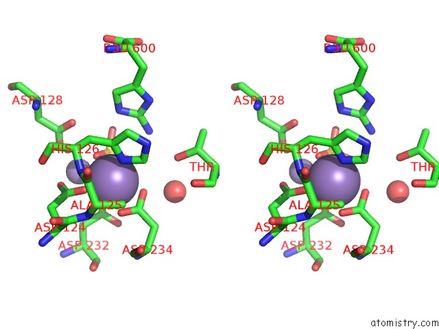
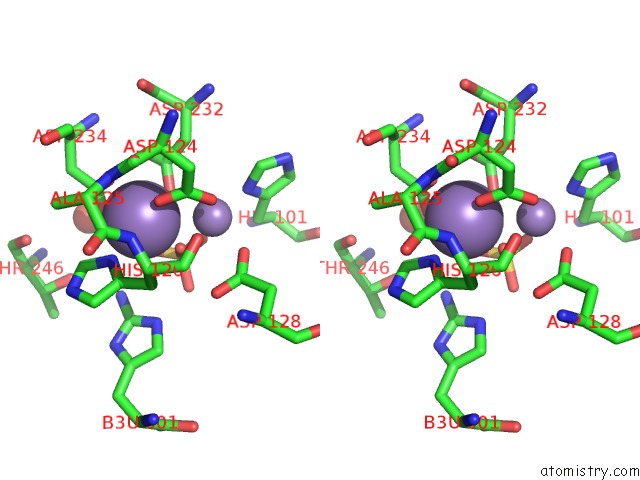
Manganese binding site 2 out of 4 in 3mfw
Go back to
Manganese binding site 2 out
of 4 in the Crystal Structure of Human Arginase I in Complex with L-2- Aminohistidine and Sulphate
Mono view
Stereo pair view
Mono view
Stereo pair view
A full contact list of Manganese with other atoms in the Mn binding
site number 2 of Crystal Structure of Human Arginase I in Complex with L-2- Aminohistidine and Sulphate within 5.0Å range:
|
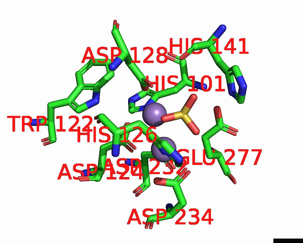
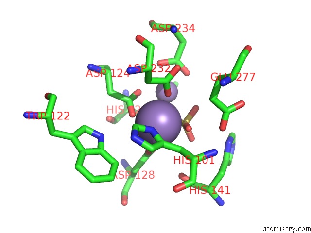
Manganese binding site 3 out of 4 in 3mfw
Go back to
Manganese binding site 3 out
of 4 in the Crystal Structure of Human Arginase I in Complex with L-2- Aminohistidine and Sulphate
Mono view
Stereo pair view
Mono view
Stereo pair view
A full contact list of Manganese with other atoms in the Mn binding
site number 3 of Crystal Structure of Human Arginase I in Complex with L-2- Aminohistidine and Sulphate within 5.0Å range:
|
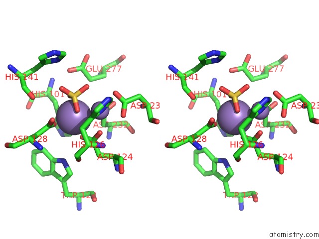
Manganese binding site 4 out of 4 in 3mfw
Go back to
Manganese binding site 4 out
of 4 in the Crystal Structure of Human Arginase I in Complex with L-2- Aminohistidine and Sulphate
Mono view
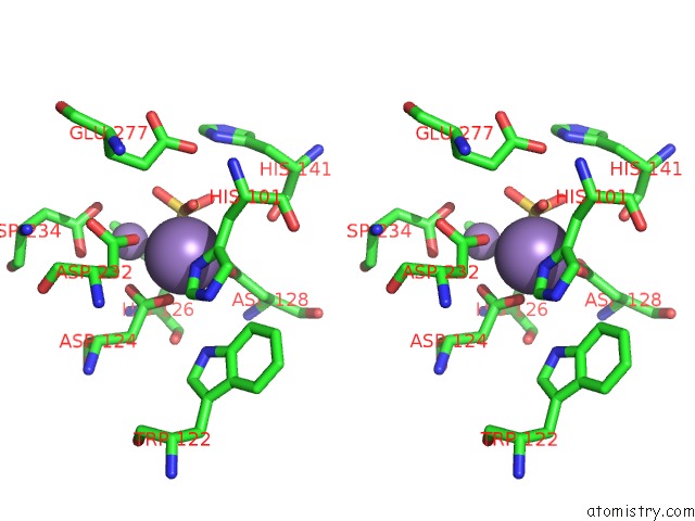
Stereo pair view
Mono view
Stereo pair view
A full contact list of Manganese with other atoms in the Mn binding
site number 4 of Crystal Structure of Human Arginase I in Complex with L-2- Aminohistidine and Sulphate within 5.0Å range:
|
Reference:
M.Ilies,
L.Di Costanzo,
M.L.North,
J.A.Scott,
D.W.Christianson.
2-Aminoimidazole Amino Acids As Inhibitors of the Binuclear Manganese Metalloenzyme Human Arginase I. J.Med.Chem. V. 53 4266 2010.
ISSN: ISSN 0022-2623
PubMed: 20441173
DOI: 10.1021/JM100306A
Page generated: Sat Oct 5 17:07:35 2024
ISSN: ISSN 0022-2623
PubMed: 20441173
DOI: 10.1021/JM100306A
Last articles
K in 4TTGK in 4UA6
K in 4UD8
K in 4U76
K in 4U75
K in 4U73
K in 4U71
K in 4U70
K in 4U6Z
K in 4TS0