Manganese »
PDB 3kky-3m0l »
3lp4 »
Manganese in PDB 3lp4: Crystal Structure of Human Arginase I in Complex with L-Lysine, 1.90A Resolution.
Enzymatic activity of Crystal Structure of Human Arginase I in Complex with L-Lysine, 1.90A Resolution.
All present enzymatic activity of Crystal Structure of Human Arginase I in Complex with L-Lysine, 1.90A Resolution.:
3.5.3.1;
3.5.3.1;
Protein crystallography data
The structure of Crystal Structure of Human Arginase I in Complex with L-Lysine, 1.90A Resolution., PDB code: 3lp4
was solved by
L.Di Costanzo,
D.W.Christianson,
with X-Ray Crystallography technique. A brief refinement statistics is given in the table below:
| Resolution Low / High (Å) | 34.20 / 1.90 |
| Space group | P 3 |
| Cell size a, b, c (Å), α, β, γ (°) | 90.729, 90.729, 69.509, 90.00, 90.00, 120.00 |
| R / Rfree (%) | 15.5 / 20.4 |
Manganese Binding Sites:
The binding sites of Manganese atom in the Crystal Structure of Human Arginase I in Complex with L-Lysine, 1.90A Resolution.
(pdb code 3lp4). This binding sites where shown within
5.0 Angstroms radius around Manganese atom.
In total 4 binding sites of Manganese where determined in the Crystal Structure of Human Arginase I in Complex with L-Lysine, 1.90A Resolution., PDB code: 3lp4:
Jump to Manganese binding site number: 1; 2; 3; 4;
In total 4 binding sites of Manganese where determined in the Crystal Structure of Human Arginase I in Complex with L-Lysine, 1.90A Resolution., PDB code: 3lp4:
Jump to Manganese binding site number: 1; 2; 3; 4;
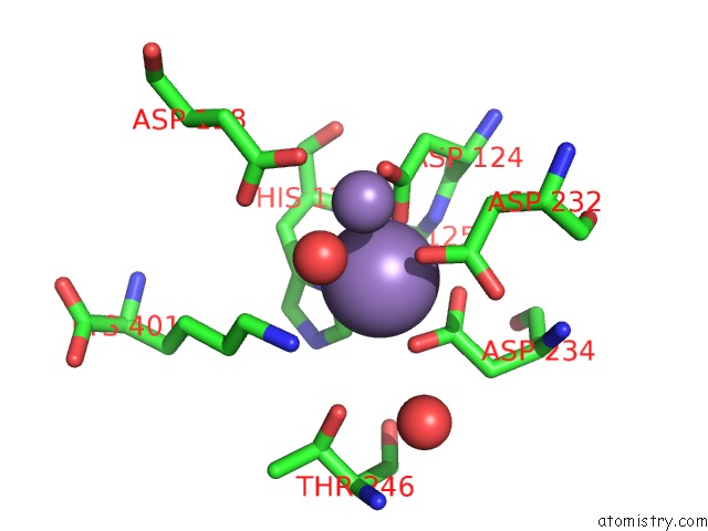
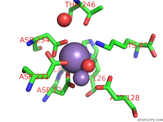
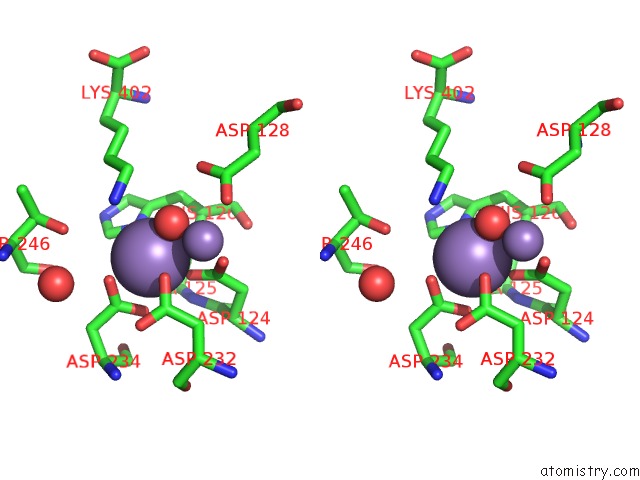
Manganese binding site 1 out of 4 in 3lp4
Go back to
Manganese binding site 1 out
of 4 in the Crystal Structure of Human Arginase I in Complex with L-Lysine, 1.90A Resolution.
Mono view
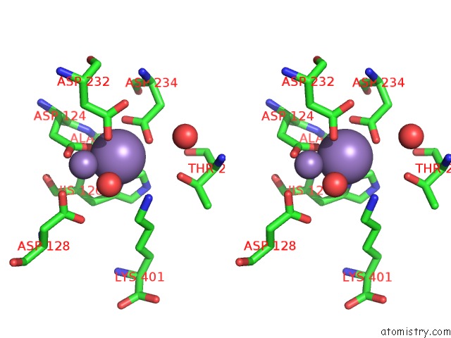
Stereo pair view
Mono view
Stereo pair view
A full contact list of Manganese with other atoms in the Mn binding
site number 1 of Crystal Structure of Human Arginase I in Complex with L-Lysine, 1.90A Resolution. within 5.0Å range:
|
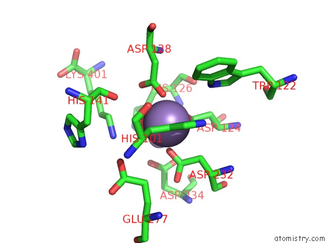
Manganese binding site 2 out of 4 in 3lp4
Go back to
Manganese binding site 2 out
of 4 in the Crystal Structure of Human Arginase I in Complex with L-Lysine, 1.90A Resolution.
Mono view
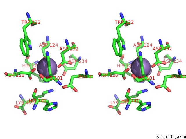
Stereo pair view
Mono view
Stereo pair view
A full contact list of Manganese with other atoms in the Mn binding
site number 2 of Crystal Structure of Human Arginase I in Complex with L-Lysine, 1.90A Resolution. within 5.0Å range:
|
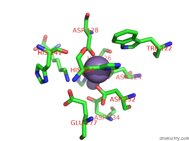
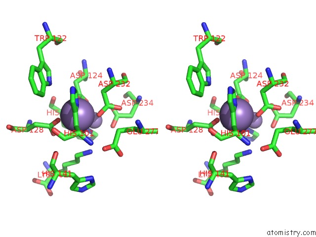
Manganese binding site 3 out of 4 in 3lp4
Go back to
Manganese binding site 3 out
of 4 in the Crystal Structure of Human Arginase I in Complex with L-Lysine, 1.90A Resolution.
Mono view
Stereo pair view
Mono view
Stereo pair view
A full contact list of Manganese with other atoms in the Mn binding
site number 3 of Crystal Structure of Human Arginase I in Complex with L-Lysine, 1.90A Resolution. within 5.0Å range:
|
Manganese binding site 4 out of 4 in 3lp4
Go back to
Manganese binding site 4 out
of 4 in the Crystal Structure of Human Arginase I in Complex with L-Lysine, 1.90A Resolution.
Mono view
Stereo pair view
Mono view
Stereo pair view
A full contact list of Manganese with other atoms in the Mn binding
site number 4 of Crystal Structure of Human Arginase I in Complex with L-Lysine, 1.90A Resolution. within 5.0Å range:
|
Reference:
L.Di Costanzo,
M.Ilies,
K.J.Thorn,
D.W.Christianson.
Inhibition of Human Arginase I By Substrate and Product Analogues. Arch.Biochem.Biophys. V. 496 101 2010.
ISSN: ISSN 0003-9861
PubMed: 20153713
DOI: 10.1016/J.ABB.2010.02.004
Page generated: Sat Oct 5 16:52:14 2024
ISSN: ISSN 0003-9861
PubMed: 20153713
DOI: 10.1016/J.ABB.2010.02.004
Last articles
I in 4PNSI in 4P4Z
I in 4P1E
I in 4P4X
I in 4P4Y
I in 4P4W
I in 4OA5
I in 4OWA
I in 4P4V
I in 4OWC