Manganese »
PDB 3hw6-3ki9 »
3kdj »
Manganese in PDB 3kdj: Complex Structure of (+)-Aba-Bound PYL1 and ABI1
Enzymatic activity of Complex Structure of (+)-Aba-Bound PYL1 and ABI1
All present enzymatic activity of Complex Structure of (+)-Aba-Bound PYL1 and ABI1:
3.1.3.16;
3.1.3.16;
Protein crystallography data
The structure of Complex Structure of (+)-Aba-Bound PYL1 and ABI1, PDB code: 3kdj
was solved by
P.Yin,
H.Fan,
Q.Hao,
X.Yuan,
N.Yan,
with X-Ray Crystallography technique. A brief refinement statistics is given in the table below:
| Resolution Low / High (Å) | 34.15 / 1.88 |
| Space group | P 21 21 21 |
| Cell size a, b, c (Å), α, β, γ (°) | 61.610, 86.788, 110.701, 90.00, 90.00, 90.00 |
| R / Rfree (%) | 20.9 / 24.6 |
Manganese Binding Sites:
The binding sites of Manganese atom in the Complex Structure of (+)-Aba-Bound PYL1 and ABI1
(pdb code 3kdj). This binding sites where shown within
5.0 Angstroms radius around Manganese atom.
In total only one binding site of Manganese was determined in the Complex Structure of (+)-Aba-Bound PYL1 and ABI1, PDB code: 3kdj:
In total only one binding site of Manganese was determined in the Complex Structure of (+)-Aba-Bound PYL1 and ABI1, PDB code: 3kdj:
Manganese binding site 1 out of 1 in 3kdj
Go back to
Manganese binding site 1 out
of 1 in the Complex Structure of (+)-Aba-Bound PYL1 and ABI1
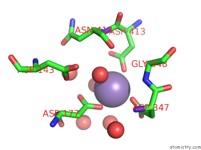
Mono view
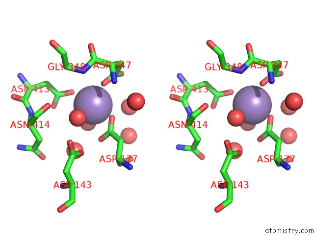
Stereo pair view
Mono view
Stereo pair view
A full contact list of Manganese with other atoms in the Mn binding
site number 1 of Complex Structure of (+)-Aba-Bound PYL1 and ABI1 within 5.0Å range:
|
Reference:
P.Yin,
H.Fan,
Q.Hao,
X.Yuan,
D.Wu,
Y.Pang,
C.Yan,
W.Li,
J.Wang,
N.Yan.
Structural Insights Into the Mechanism of Abscisic Acid Signaling By Pyl Proteins Nat.Struct.Mol.Biol. V. 16 1230 2009.
ISSN: ISSN 1545-9993
PubMed: 19893533
DOI: 10.1038/NSMB.1730
Page generated: Sat Oct 5 16:43:58 2024
ISSN: ISSN 1545-9993
PubMed: 19893533
DOI: 10.1038/NSMB.1730
Last articles
Mg in 5NS4Mg in 5NUG
Mg in 5NV1
Mg in 5NPU
Mg in 5NRT
Mg in 5NRO
Mg in 5NRI
Mg in 5NRH
Mg in 5NQU
Mg in 5NQT