Manganese »
PDB 3g0z-3hvq »
3hmk »
Manganese in PDB 3hmk: Crystal Structure of Serine Racemase
Enzymatic activity of Crystal Structure of Serine Racemase
All present enzymatic activity of Crystal Structure of Serine Racemase:
5.1.1.18;
5.1.1.18;
Protein crystallography data
The structure of Crystal Structure of Serine Racemase, PDB code: 3hmk
was solved by
M.A.Smith,
J.Barker,
V.Mack,
A.Ebneth,
B.Felicetti,
M.Woods,
with X-Ray Crystallography technique. A brief refinement statistics is given in the table below:
| Resolution Low / High (Å) | 41.10 / 2.10 |
| Space group | P 21 21 21 |
| Cell size a, b, c (Å), α, β, γ (°) | 48.261, 102.960, 120.923, 90.00, 90.00, 90.00 |
| R / Rfree (%) | 24.1 / 28.9 |
Manganese Binding Sites:
The binding sites of Manganese atom in the Crystal Structure of Serine Racemase
(pdb code 3hmk). This binding sites where shown within
5.0 Angstroms radius around Manganese atom.
In total 2 binding sites of Manganese where determined in the Crystal Structure of Serine Racemase, PDB code: 3hmk:
Jump to Manganese binding site number: 1; 2;
In total 2 binding sites of Manganese where determined in the Crystal Structure of Serine Racemase, PDB code: 3hmk:
Jump to Manganese binding site number: 1; 2;
Manganese binding site 1 out of 2 in 3hmk
Go back to
Manganese binding site 1 out
of 2 in the Crystal Structure of Serine Racemase
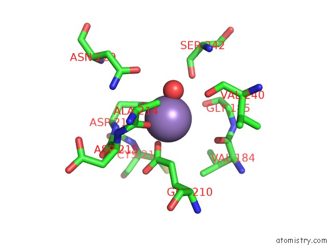
Mono view
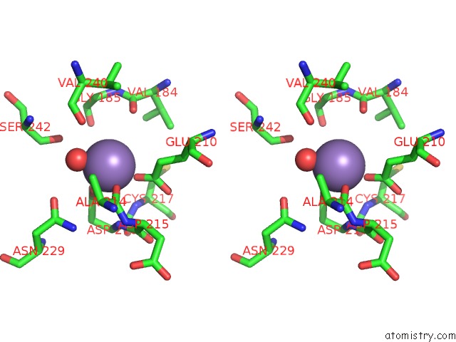
Stereo pair view
Mono view
Stereo pair view
A full contact list of Manganese with other atoms in the Mn binding
site number 1 of Crystal Structure of Serine Racemase within 5.0Å range:
|
Manganese binding site 2 out of 2 in 3hmk
Go back to
Manganese binding site 2 out
of 2 in the Crystal Structure of Serine Racemase
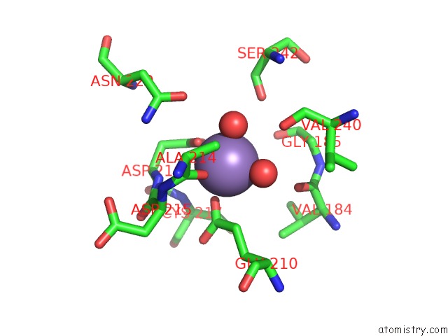
Mono view
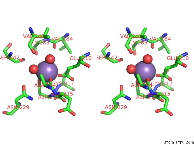
Stereo pair view
Mono view
Stereo pair view
A full contact list of Manganese with other atoms in the Mn binding
site number 2 of Crystal Structure of Serine Racemase within 5.0Å range:
|
Reference:
M.A.Smith,
V.Mack,
A.Ebneth,
I.Moraes,
B.Felicetti,
M.Wood,
D.Schonfeld,
O.Mather,
A.Cesura,
J.Barker.
The Structure of Mammalian Serine Racemase: Evidence For Conformational Changes Upon Inhibitor Binding. J.Biol.Chem. V. 285 12873 2010.
ISSN: ISSN 0021-9258
PubMed: 20106978
DOI: 10.1074/JBC.M109.050062
Page generated: Sat Aug 16 11:55:10 2025
ISSN: ISSN 0021-9258
PubMed: 20106978
DOI: 10.1074/JBC.M109.050062
Last articles
Na in 6L4PNa in 6L46
Na in 6L19
Na in 6L18
Na in 6KW1
Na in 6KXH
Na in 6KTR
Na in 6KV0
Na in 6KUM
Na in 6KTW