Manganese »
PDB 3g0z-3hvq »
3hix »
Manganese in PDB 3hix: Crystal Structure of the RHODANESE_3 Like Domain From Anabaena Sp ALR3790 Protein. Northeast Structural Genomics Consortium Target NSR437I
Protein crystallography data
The structure of Crystal Structure of the RHODANESE_3 Like Domain From Anabaena Sp ALR3790 Protein. Northeast Structural Genomics Consortium Target NSR437I, PDB code: 3hix
was solved by
S.Vorobiev,
Y.Chen,
F.Forouhar,
M.Maglaqui,
C.Ciccosanti,
L.Mao,
R.Xiao,
T.B.Acton,
G.T.Montelione,
L.Tong,
J.F.Hunt,
Northeaststructural Genomics Consortium (Nesg),
with X-Ray Crystallography technique. A brief refinement statistics is given in the table below:
| Resolution Low / High (Å) | 34.61 / 1.92 |
| Space group | P 21 21 2 |
| Cell size a, b, c (Å), α, β, γ (°) | 62.306, 105.479, 41.615, 90.00, 90.00, 90.00 |
| R / Rfree (%) | 21.4 / 23 |
Manganese Binding Sites:
The binding sites of Manganese atom in the Crystal Structure of the RHODANESE_3 Like Domain From Anabaena Sp ALR3790 Protein. Northeast Structural Genomics Consortium Target NSR437I
(pdb code 3hix). This binding sites where shown within
5.0 Angstroms radius around Manganese atom.
In total 5 binding sites of Manganese where determined in the Crystal Structure of the RHODANESE_3 Like Domain From Anabaena Sp ALR3790 Protein. Northeast Structural Genomics Consortium Target NSR437I, PDB code: 3hix:
Jump to Manganese binding site number: 1; 2; 3; 4; 5;
In total 5 binding sites of Manganese where determined in the Crystal Structure of the RHODANESE_3 Like Domain From Anabaena Sp ALR3790 Protein. Northeast Structural Genomics Consortium Target NSR437I, PDB code: 3hix:
Jump to Manganese binding site number: 1; 2; 3; 4; 5;
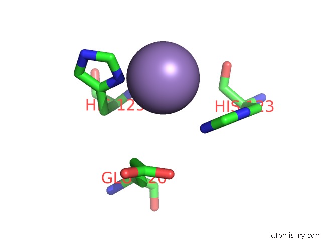
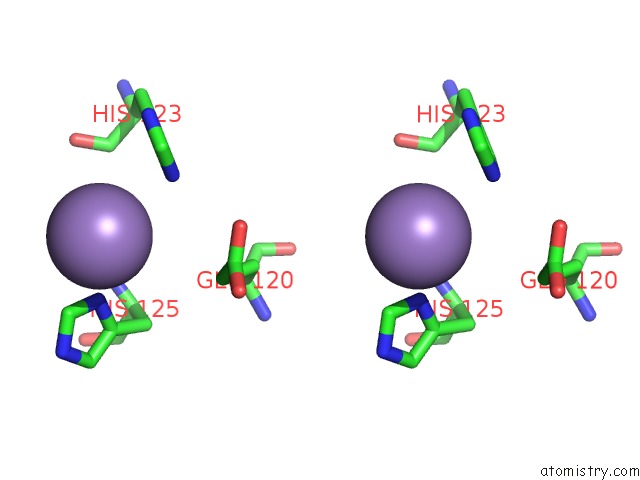
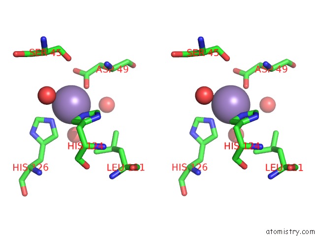
Manganese binding site 1 out of 5 in 3hix
Go back to
Manganese binding site 1 out
of 5 in the Crystal Structure of the RHODANESE_3 Like Domain From Anabaena Sp ALR3790 Protein. Northeast Structural Genomics Consortium Target NSR437I
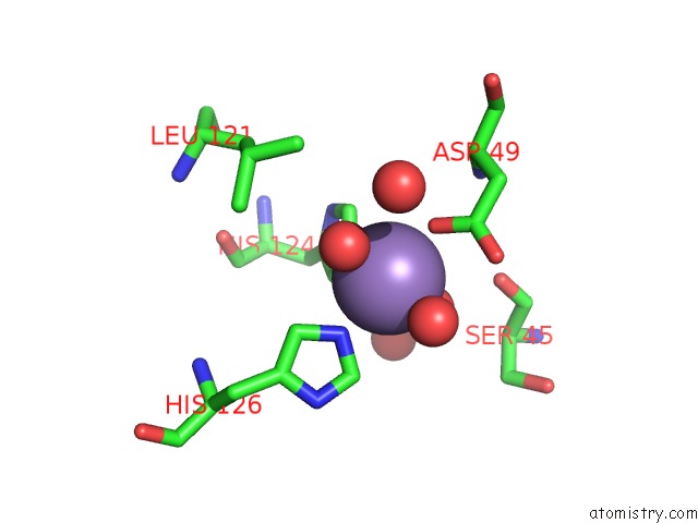
Mono view
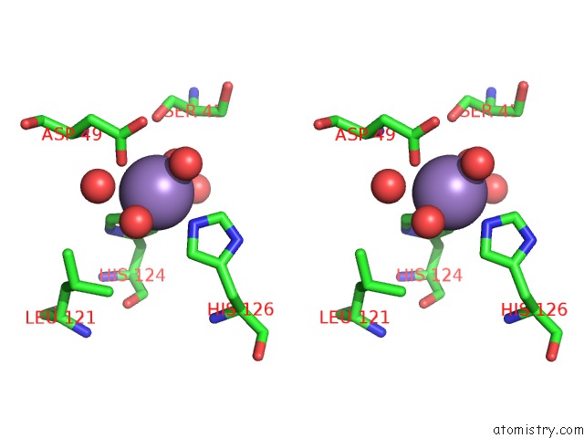
Stereo pair view
Mono view
Stereo pair view
A full contact list of Manganese with other atoms in the Mn binding
site number 1 of Crystal Structure of the RHODANESE_3 Like Domain From Anabaena Sp ALR3790 Protein. Northeast Structural Genomics Consortium Target NSR437I within 5.0Å range:
|
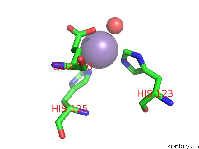
Manganese binding site 2 out of 5 in 3hix
Go back to
Manganese binding site 2 out
of 5 in the Crystal Structure of the RHODANESE_3 Like Domain From Anabaena Sp ALR3790 Protein. Northeast Structural Genomics Consortium Target NSR437I
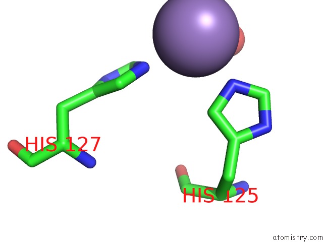
Mono view
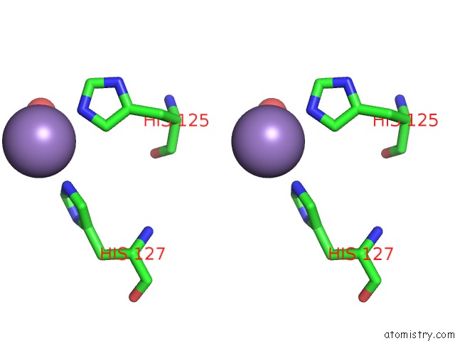
Stereo pair view
Mono view
Stereo pair view
A full contact list of Manganese with other atoms in the Mn binding
site number 2 of Crystal Structure of the RHODANESE_3 Like Domain From Anabaena Sp ALR3790 Protein. Northeast Structural Genomics Consortium Target NSR437I within 5.0Å range:
|
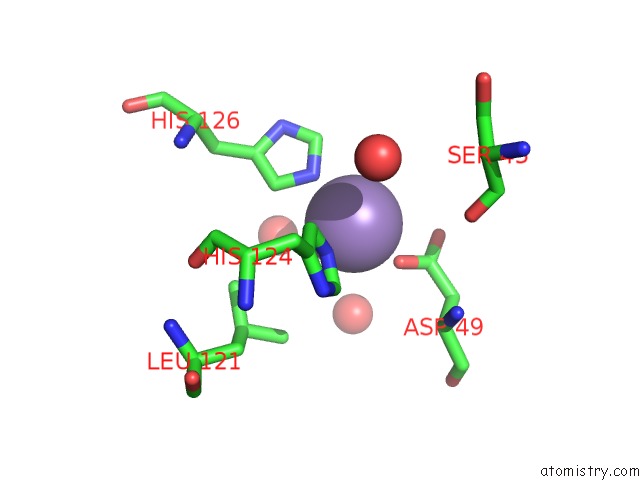
Manganese binding site 3 out of 5 in 3hix
Go back to
Manganese binding site 3 out
of 5 in the Crystal Structure of the RHODANESE_3 Like Domain From Anabaena Sp ALR3790 Protein. Northeast Structural Genomics Consortium Target NSR437I
Mono view
Stereo pair view
Mono view
Stereo pair view
A full contact list of Manganese with other atoms in the Mn binding
site number 3 of Crystal Structure of the RHODANESE_3 Like Domain From Anabaena Sp ALR3790 Protein. Northeast Structural Genomics Consortium Target NSR437I within 5.0Å range:
|
Manganese binding site 4 out of 5 in 3hix
Go back to
Manganese binding site 4 out
of 5 in the Crystal Structure of the RHODANESE_3 Like Domain From Anabaena Sp ALR3790 Protein. Northeast Structural Genomics Consortium Target NSR437I
Mono view

Stereo pair view
Mono view
Stereo pair view
A full contact list of Manganese with other atoms in the Mn binding
site number 4 of Crystal Structure of the RHODANESE_3 Like Domain From Anabaena Sp ALR3790 Protein. Northeast Structural Genomics Consortium Target NSR437I within 5.0Å range:
|
Manganese binding site 5 out of 5 in 3hix
Go back to
Manganese binding site 5 out
of 5 in the Crystal Structure of the RHODANESE_3 Like Domain From Anabaena Sp ALR3790 Protein. Northeast Structural Genomics Consortium Target NSR437I
Mono view
Stereo pair view
Mono view
Stereo pair view
A full contact list of Manganese with other atoms in the Mn binding
site number 5 of Crystal Structure of the RHODANESE_3 Like Domain From Anabaena Sp ALR3790 Protein. Northeast Structural Genomics Consortium Target NSR437I within 5.0Å range:
|
Reference:
S.Vorobiev,
Y.Chen,
F.Forouhar,
M.Maglaqui,
C.Ciccosanti,
L.Mao,
R.Xiao,
T.B.Acton,
G.T.Montelione,
L.Tong,
J.F.Hunt.
Crystal Structure of the RHODANESE_3 Like Domain From Anabaena Sp ALR3790 Protein. To Be Published.
Page generated: Sat Aug 16 11:55:08 2025
Last articles
Na in 3MMONa in 3MO9
Na in 3MO6
Na in 3MO3
Na in 3MNZ
Na in 3MNX
Na in 3MNS
Na in 3MNK
Na in 3MNJ
Na in 3MNI