Manganese »
PDB 3ebc-3g0a »
3fcm »
Manganese in PDB 3fcm: Crystal Structure of A Nudix Hydrolase From Clostridium Perfringens
Protein crystallography data
The structure of Crystal Structure of A Nudix Hydrolase From Clostridium Perfringens, PDB code: 3fcm
was solved by
K.Palani,
S.K.Burley,
S.Swaninathan,
New York Sgx Research Center Forstructural Genomics (Nysgxrc),
with X-Ray Crystallography technique. A brief refinement statistics is given in the table below:
| Resolution Low / High (Å) | 39.48 / 2.20 |
| Space group | P 21 21 2 |
| Cell size a, b, c (Å), α, β, γ (°) | 80.395, 132.459, 41.361, 90.00, 90.00, 90.00 |
| R / Rfree (%) | 22.9 / 28 |
Manganese Binding Sites:
The binding sites of Manganese atom in the Crystal Structure of A Nudix Hydrolase From Clostridium Perfringens
(pdb code 3fcm). This binding sites where shown within
5.0 Angstroms radius around Manganese atom.
In total 2 binding sites of Manganese where determined in the Crystal Structure of A Nudix Hydrolase From Clostridium Perfringens, PDB code: 3fcm:
Jump to Manganese binding site number: 1; 2;
In total 2 binding sites of Manganese where determined in the Crystal Structure of A Nudix Hydrolase From Clostridium Perfringens, PDB code: 3fcm:
Jump to Manganese binding site number: 1; 2;
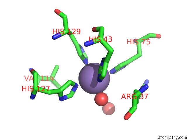
Manganese binding site 1 out of 2 in 3fcm
Go back to
Manganese binding site 1 out
of 2 in the Crystal Structure of A Nudix Hydrolase From Clostridium Perfringens
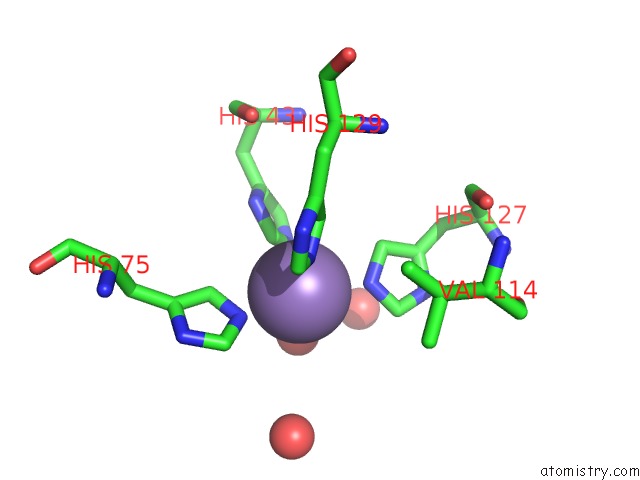
Mono view
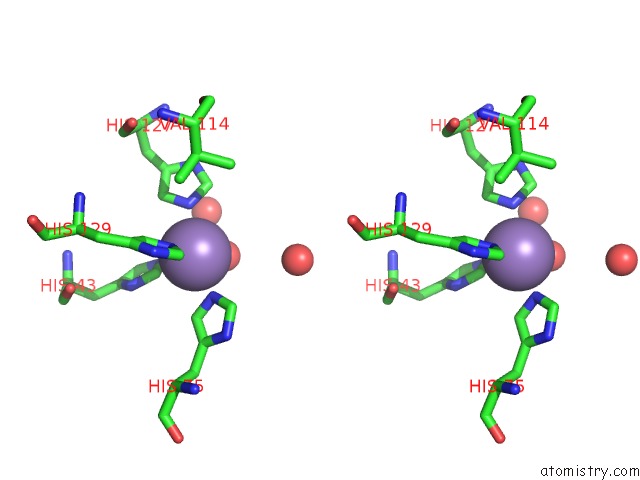
Stereo pair view
Mono view
Stereo pair view
A full contact list of Manganese with other atoms in the Mn binding
site number 1 of Crystal Structure of A Nudix Hydrolase From Clostridium Perfringens within 5.0Å range:
|
Manganese binding site 2 out of 2 in 3fcm
Go back to
Manganese binding site 2 out
of 2 in the Crystal Structure of A Nudix Hydrolase From Clostridium Perfringens
Mono view

Stereo pair view
Mono view
Stereo pair view
A full contact list of Manganese with other atoms in the Mn binding
site number 2 of Crystal Structure of A Nudix Hydrolase From Clostridium Perfringens within 5.0Å range:
|
Reference:
K.Palani,
S.K.Burley,
S.Swaminathan.
Crystal Structure of A Nudix Hydrolase From Clostridium Perfringens To Be Published.
Page generated: Sat Oct 5 16:16:00 2024
Last articles
Mg in 1SO2Mg in 1SO5
Mg in 1SO4
Mg in 1SO3
Mg in 1SNF
Mg in 1SLH
Mg in 1SL2
Mg in 1SL5
Mg in 1SKR
Mg in 1SL0