Manganese »
PDB 3ebc-3g0a »
3ee4 »
Manganese in PDB 3ee4: R2-Like Ligand Binding Mn/Fe Oxidase From M. Tuberculosis
Enzymatic activity of R2-Like Ligand Binding Mn/Fe Oxidase From M. Tuberculosis
All present enzymatic activity of R2-Like Ligand Binding Mn/Fe Oxidase From M. Tuberculosis:
1.17.4.1;
1.17.4.1;
Protein crystallography data
The structure of R2-Like Ligand Binding Mn/Fe Oxidase From M. Tuberculosis, PDB code: 3ee4
was solved by
C.S.Andersson,
T.A.Jones,
M.Hogbom,
with X-Ray Crystallography technique. A brief refinement statistics is given in the table below:
| Resolution Low / High (Å) | 36.85 / 1.90 |
| Space group | P 32 2 1 |
| Cell size a, b, c (Å), α, β, γ (°) | 54.565, 54.565, 176.651, 90.00, 90.00, 120.00 |
| R / Rfree (%) | 14.9 / 17.7 |
Other elements in 3ee4:
The structure of R2-Like Ligand Binding Mn/Fe Oxidase From M. Tuberculosis also contains other interesting chemical elements:
| Iron | (Fe) | 1 atom |
Manganese Binding Sites:
The binding sites of Manganese atom in the R2-Like Ligand Binding Mn/Fe Oxidase From M. Tuberculosis
(pdb code 3ee4). This binding sites where shown within
5.0 Angstroms radius around Manganese atom.
In total only one binding site of Manganese was determined in the R2-Like Ligand Binding Mn/Fe Oxidase From M. Tuberculosis, PDB code: 3ee4:
In total only one binding site of Manganese was determined in the R2-Like Ligand Binding Mn/Fe Oxidase From M. Tuberculosis, PDB code: 3ee4:
Manganese binding site 1 out of 1 in 3ee4
Go back to
Manganese binding site 1 out
of 1 in the R2-Like Ligand Binding Mn/Fe Oxidase From M. Tuberculosis
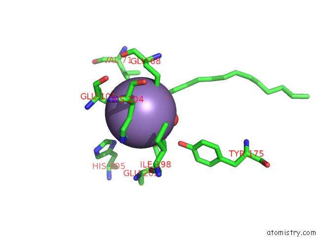
Mono view
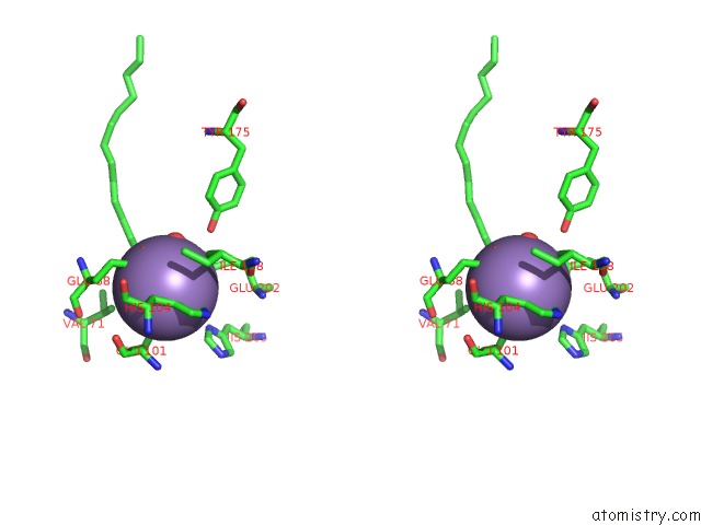
Stereo pair view
Mono view
Stereo pair view
A full contact list of Manganese with other atoms in the Mn binding
site number 1 of R2-Like Ligand Binding Mn/Fe Oxidase From M. Tuberculosis within 5.0Å range:
|
Reference:
C.S.Andersson,
C.S.Andersson,
T.A.Jones,
M.Hogbom.
N/A N/A.
ISSN: ISSN 0027-8424
PubMed: 19321420
DOI: 10.1073/PNAS.0812971106
Page generated: Sat Aug 16 11:41:27 2025
ISSN: ISSN 0027-8424
PubMed: 19321420
DOI: 10.1073/PNAS.0812971106
Last articles
Yb in 3FTXYb in 3FTY
Yb in 3FTV
Yb in 3FTW
Yb in 3FHE
Yb in 3FTU
Yb in 3FH8
Yb in 3FTS
Yb in 3FH7
Yb in 3FH5