Manganese »
PDB 3auz-3c5m »
3bza »
Manganese in PDB 3bza: Structure of Mn-Substituted Homoprotocatechuate 2,3-Dioxygenase From B.Fuscum at 1.7 Ang Resolution
Enzymatic activity of Structure of Mn-Substituted Homoprotocatechuate 2,3-Dioxygenase From B.Fuscum at 1.7 Ang Resolution
All present enzymatic activity of Structure of Mn-Substituted Homoprotocatechuate 2,3-Dioxygenase From B.Fuscum at 1.7 Ang Resolution:
1.13.11.15;
1.13.11.15;
Protein crystallography data
The structure of Structure of Mn-Substituted Homoprotocatechuate 2,3-Dioxygenase From B.Fuscum at 1.7 Ang Resolution, PDB code: 3bza
was solved by
E.G.Kovaleva,
J.D.Lipscomb,
with X-Ray Crystallography technique. A brief refinement statistics is given in the table below:
| Resolution Low / High (Å) | 28.82 / 1.70 |
| Space group | P 21 21 2 |
| Cell size a, b, c (Å), α, β, γ (°) | 110.502, 152.191, 96.278, 90.00, 90.00, 90.00 |
| R / Rfree (%) | 17 / 19.7 |
Other elements in 3bza:
The structure of Structure of Mn-Substituted Homoprotocatechuate 2,3-Dioxygenase From B.Fuscum at 1.7 Ang Resolution also contains other interesting chemical elements:
| Chlorine | (Cl) | 4 atoms |
| Calcium | (Ca) | 1 atom |
Manganese Binding Sites:
The binding sites of Manganese atom in the Structure of Mn-Substituted Homoprotocatechuate 2,3-Dioxygenase From B.Fuscum at 1.7 Ang Resolution
(pdb code 3bza). This binding sites where shown within
5.0 Angstroms radius around Manganese atom.
In total 4 binding sites of Manganese where determined in the Structure of Mn-Substituted Homoprotocatechuate 2,3-Dioxygenase From B.Fuscum at 1.7 Ang Resolution, PDB code: 3bza:
Jump to Manganese binding site number: 1; 2; 3; 4;
In total 4 binding sites of Manganese where determined in the Structure of Mn-Substituted Homoprotocatechuate 2,3-Dioxygenase From B.Fuscum at 1.7 Ang Resolution, PDB code: 3bza:
Jump to Manganese binding site number: 1; 2; 3; 4;
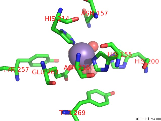
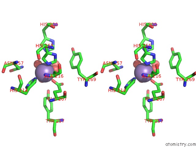
Manganese binding site 1 out of 4 in 3bza
Go back to
Manganese binding site 1 out
of 4 in the Structure of Mn-Substituted Homoprotocatechuate 2,3-Dioxygenase From B.Fuscum at 1.7 Ang Resolution
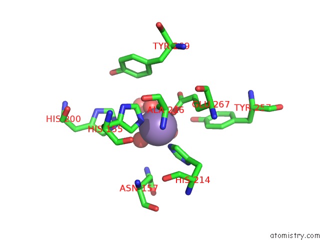
Mono view
Stereo pair view
Mono view
Stereo pair view
A full contact list of Manganese with other atoms in the Mn binding
site number 1 of Structure of Mn-Substituted Homoprotocatechuate 2,3-Dioxygenase From B.Fuscum at 1.7 Ang Resolution within 5.0Å range:
|
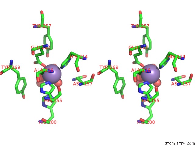
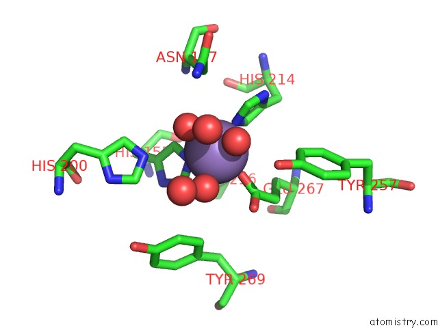
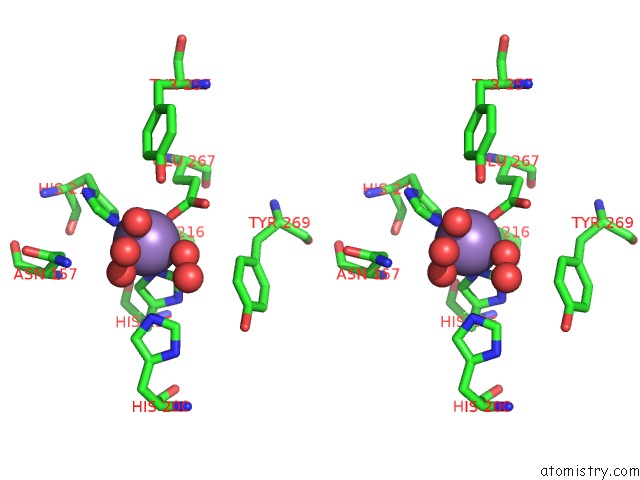
Manganese binding site 2 out of 4 in 3bza
Go back to
Manganese binding site 2 out
of 4 in the Structure of Mn-Substituted Homoprotocatechuate 2,3-Dioxygenase From B.Fuscum at 1.7 Ang Resolution
Mono view
Stereo pair view
Mono view
Stereo pair view
A full contact list of Manganese with other atoms in the Mn binding
site number 2 of Structure of Mn-Substituted Homoprotocatechuate 2,3-Dioxygenase From B.Fuscum at 1.7 Ang Resolution within 5.0Å range:
|
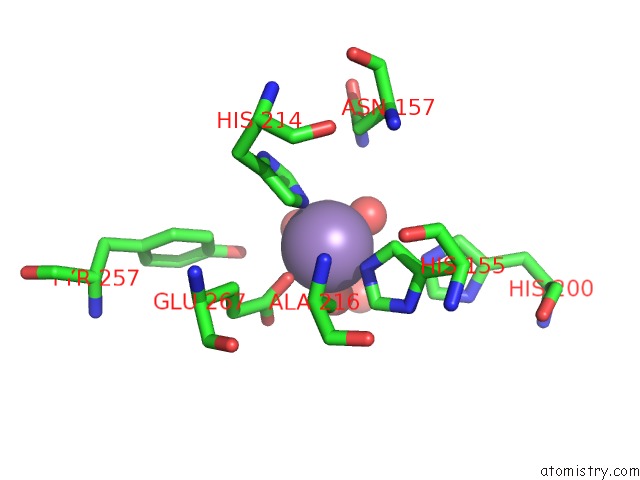
Manganese binding site 3 out of 4 in 3bza
Go back to
Manganese binding site 3 out
of 4 in the Structure of Mn-Substituted Homoprotocatechuate 2,3-Dioxygenase From B.Fuscum at 1.7 Ang Resolution
Mono view
Stereo pair view
Mono view
Stereo pair view
A full contact list of Manganese with other atoms in the Mn binding
site number 3 of Structure of Mn-Substituted Homoprotocatechuate 2,3-Dioxygenase From B.Fuscum at 1.7 Ang Resolution within 5.0Å range:
|
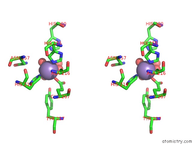
Manganese binding site 4 out of 4 in 3bza
Go back to
Manganese binding site 4 out
of 4 in the Structure of Mn-Substituted Homoprotocatechuate 2,3-Dioxygenase From B.Fuscum at 1.7 Ang Resolution
Mono view
Stereo pair view
Mono view
Stereo pair view
A full contact list of Manganese with other atoms in the Mn binding
site number 4 of Structure of Mn-Substituted Homoprotocatechuate 2,3-Dioxygenase From B.Fuscum at 1.7 Ang Resolution within 5.0Å range:
|
Reference:
J.P.Emerson,
E.G.Kovaleva,
E.R.Farquhar,
J.D.Lipscomb,
L.Que.
Swapping Metals in Fe- and Mn-Dependent Dioxygenases: Evidence For Oxygen Activation Without A Change in Metal Redox State. Proc.Natl.Acad.Sci.Usa V. 105 7347 2008.
ISSN: ISSN 0027-8424
PubMed: 18492808
DOI: 10.1073/PNAS.0711179105
Page generated: Sat Oct 5 15:59:20 2024
ISSN: ISSN 0027-8424
PubMed: 18492808
DOI: 10.1073/PNAS.0711179105
Last articles
K in 4EVYK in 4EOU
K in 4ETM
K in 4ESK
K in 4ES8
K in 4ERT
K in 4ERD
K in 4ENC
K in 4EK1
K in 4ENB