Manganese »
PDB 2ypq-3au8 »
2zxp »
Manganese in PDB 2zxp: Crystal Structure of Recj in Complex with MN2+ From Thermus Thermophilus HB8
Protein crystallography data
The structure of Crystal Structure of Recj in Complex with MN2+ From Thermus Thermophilus HB8, PDB code: 2zxp
was solved by
T.Wakamatsu,
Y.Kitamura,
N.Nakagawa,
R.Masui,
S.Kuramitsu,
with X-Ray Crystallography technique. A brief refinement statistics is given in the table below:
| Resolution Low / High (Å) | 41.56 / 2.30 |
| Space group | P 43 21 2 |
| Cell size a, b, c (Å), α, β, γ (°) | 83.127, 83.127, 249.937, 90.00, 90.00, 90.00 |
| R / Rfree (%) | 23.2 / 28.1 |
Manganese Binding Sites:
The binding sites of Manganese atom in the Crystal Structure of Recj in Complex with MN2+ From Thermus Thermophilus HB8
(pdb code 2zxp). This binding sites where shown within
5.0 Angstroms radius around Manganese atom.
In total 2 binding sites of Manganese where determined in the Crystal Structure of Recj in Complex with MN2+ From Thermus Thermophilus HB8, PDB code: 2zxp:
Jump to Manganese binding site number: 1; 2;
In total 2 binding sites of Manganese where determined in the Crystal Structure of Recj in Complex with MN2+ From Thermus Thermophilus HB8, PDB code: 2zxp:
Jump to Manganese binding site number: 1; 2;
Manganese binding site 1 out of 2 in 2zxp
Go back to
Manganese binding site 1 out
of 2 in the Crystal Structure of Recj in Complex with MN2+ From Thermus Thermophilus HB8
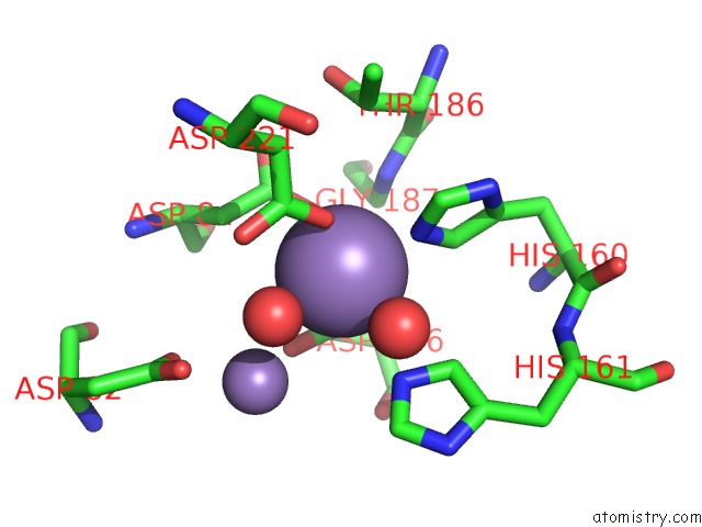
Mono view
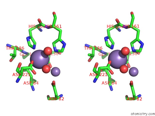
Stereo pair view
Mono view
Stereo pair view
A full contact list of Manganese with other atoms in the Mn binding
site number 1 of Crystal Structure of Recj in Complex with MN2+ From Thermus Thermophilus HB8 within 5.0Å range:
|
Manganese binding site 2 out of 2 in 2zxp
Go back to
Manganese binding site 2 out
of 2 in the Crystal Structure of Recj in Complex with MN2+ From Thermus Thermophilus HB8
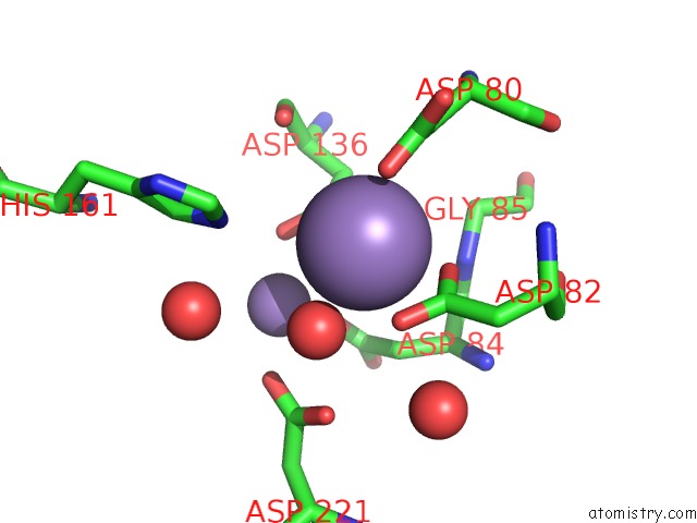
Mono view
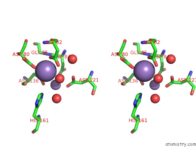
Stereo pair view
Mono view
Stereo pair view
A full contact list of Manganese with other atoms in the Mn binding
site number 2 of Crystal Structure of Recj in Complex with MN2+ From Thermus Thermophilus HB8 within 5.0Å range:
|
Reference:
T.Wakamatsu,
Y.Kitamura,
Y.Kotera,
N.Nakagawa,
S.Kuramitsu,
R.Masui.
Structure of Recj Exonuclease Defines Its Specificity For Single-Stranded Dna J.Biol.Chem. V. 285 9762 2010.
ISSN: ISSN 0021-9258
PubMed: 20129927
DOI: 10.1074/JBC.M109.096487
Page generated: Sat Aug 16 11:16:41 2025
ISSN: ISSN 0021-9258
PubMed: 20129927
DOI: 10.1074/JBC.M109.096487
Last articles
Na in 1T3MNa in 1T3P
Na in 1T2X
Na in 1SUZ
Na in 1SVY
Na in 1SVE
Na in 1SUP
Na in 1SU3
Na in 1SU4
Na in 1SUE