Manganese »
PDB 2qjc-2v3y »
2sba »
Manganese in PDB 2sba: Soybean Agglutinin Complexed with 2,6-Pentasaccharide
Protein crystallography data
The structure of Soybean Agglutinin Complexed with 2,6-Pentasaccharide, PDB code: 2sba
was solved by
A.Dessen,
D.Gupta,
S.Sabesan,
C.F.Brewer,
J.C.Sacchettini,
with X-Ray Crystallography technique. A brief refinement statistics is given in the table below:
| Resolution Low / High (Å) | 90.00 / 2.60 |
| Space group | P 64 2 2 |
| Cell size a, b, c (Å), α, β, γ (°) | 144.900, 144.900, 109.400, 90.00, 90.00, 120.00 |
| R / Rfree (%) | n/a / n/a |
Other elements in 2sba:
The structure of Soybean Agglutinin Complexed with 2,6-Pentasaccharide also contains other interesting chemical elements:
| Calcium | (Ca) | 1 atom |
Manganese Binding Sites:
The binding sites of Manganese atom in the Soybean Agglutinin Complexed with 2,6-Pentasaccharide
(pdb code 2sba). This binding sites where shown within
5.0 Angstroms radius around Manganese atom.
In total only one binding site of Manganese was determined in the Soybean Agglutinin Complexed with 2,6-Pentasaccharide, PDB code: 2sba:
In total only one binding site of Manganese was determined in the Soybean Agglutinin Complexed with 2,6-Pentasaccharide, PDB code: 2sba:
Manganese binding site 1 out of 1 in 2sba
Go back to
Manganese binding site 1 out
of 1 in the Soybean Agglutinin Complexed with 2,6-Pentasaccharide
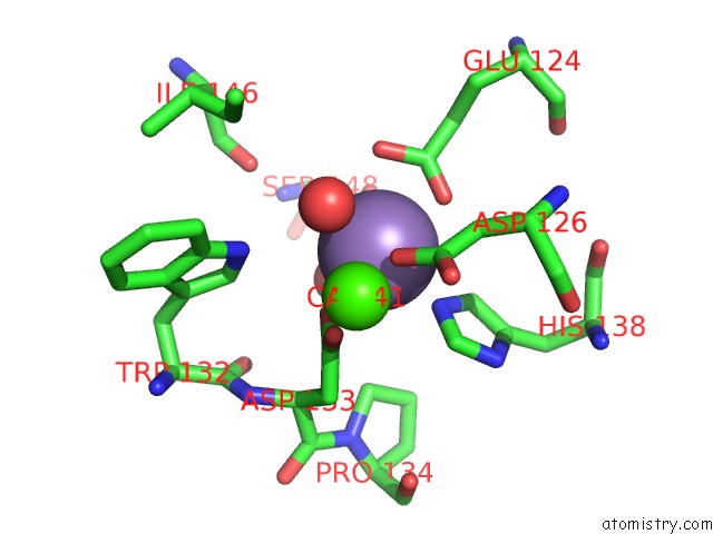
Mono view
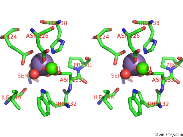
Stereo pair view
Mono view
Stereo pair view
A full contact list of Manganese with other atoms in the Mn binding
site number 1 of Soybean Agglutinin Complexed with 2,6-Pentasaccharide within 5.0Å range:
|
Reference:
A.Dessen,
D.Gupta,
S.Sabesan,
C.F.Brewer,
J.C.Sacchettini.
X-Ray Crystal Structure of the Soybean Agglutinin Cross-Linked with A Biantennary Analog of the Blood Group I Carbohydrate Antigen. Biochemistry V. 34 4933 1995.
ISSN: ISSN 0006-2960
PubMed: 7711015
DOI: 10.1021/BI00015A004
Page generated: Sat Oct 5 15:10:42 2024
ISSN: ISSN 0006-2960
PubMed: 7711015
DOI: 10.1021/BI00015A004
Last articles
Mg in 1S5LMg in 1S8F
Mg in 1S83
Mg in 1S77
Mg in 1S76
Mg in 1S4E
Mg in 1S6P
Mg in 1S6H
Mg in 1S5J
Mg in 1S5G