Manganese »
PDB 2pal-2qgi »
2qcs »
Manganese in PDB 2qcs: A Complex Structure Between the Catalytic and Regulatory Subunit of Protein Kinase A That Represents the Inhibited State
Enzymatic activity of A Complex Structure Between the Catalytic and Regulatory Subunit of Protein Kinase A That Represents the Inhibited State
All present enzymatic activity of A Complex Structure Between the Catalytic and Regulatory Subunit of Protein Kinase A That Represents the Inhibited State:
2.7.11.11;
2.7.11.11;
Protein crystallography data
The structure of A Complex Structure Between the Catalytic and Regulatory Subunit of Protein Kinase A That Represents the Inhibited State, PDB code: 2qcs
was solved by
C.Kim,
C.Y.Cheng,
A.S.Saldanha,
S.S.Taylor,
with X-Ray Crystallography technique. A brief refinement statistics is given in the table below:
| Resolution Low / High (Å) | 50.00 / 2.20 |
| Space group | P 32 2 1 |
| Cell size a, b, c (Å), α, β, γ (°) | 125.809, 125.809, 140.941, 90.00, 90.00, 120.00 |
| R / Rfree (%) | 19.2 / 22.5 |
Manganese Binding Sites:
The binding sites of Manganese atom in the A Complex Structure Between the Catalytic and Regulatory Subunit of Protein Kinase A That Represents the Inhibited State
(pdb code 2qcs). This binding sites where shown within
5.0 Angstroms radius around Manganese atom.
In total 2 binding sites of Manganese where determined in the A Complex Structure Between the Catalytic and Regulatory Subunit of Protein Kinase A That Represents the Inhibited State, PDB code: 2qcs:
Jump to Manganese binding site number: 1; 2;
In total 2 binding sites of Manganese where determined in the A Complex Structure Between the Catalytic and Regulatory Subunit of Protein Kinase A That Represents the Inhibited State, PDB code: 2qcs:
Jump to Manganese binding site number: 1; 2;
Manganese binding site 1 out of 2 in 2qcs
Go back to
Manganese binding site 1 out
of 2 in the A Complex Structure Between the Catalytic and Regulatory Subunit of Protein Kinase A That Represents the Inhibited State
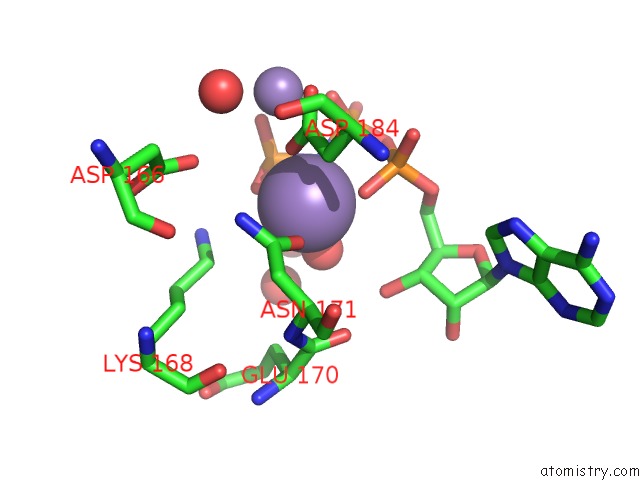
Mono view
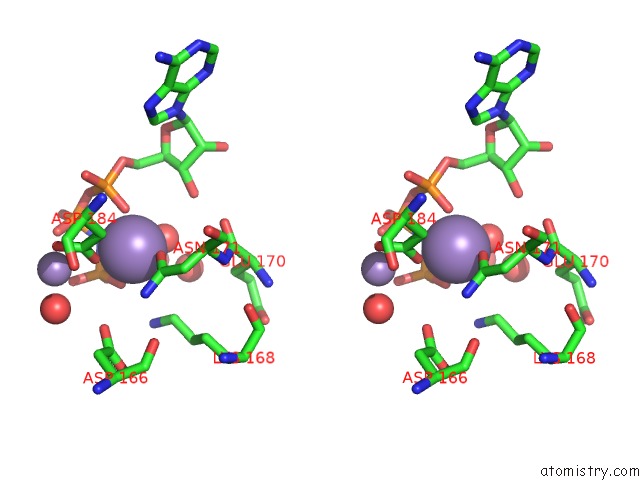
Stereo pair view
Mono view
Stereo pair view
A full contact list of Manganese with other atoms in the Mn binding
site number 1 of A Complex Structure Between the Catalytic and Regulatory Subunit of Protein Kinase A That Represents the Inhibited State within 5.0Å range:
|
Manganese binding site 2 out of 2 in 2qcs
Go back to
Manganese binding site 2 out
of 2 in the A Complex Structure Between the Catalytic and Regulatory Subunit of Protein Kinase A That Represents the Inhibited State
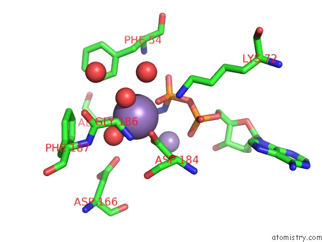
Mono view
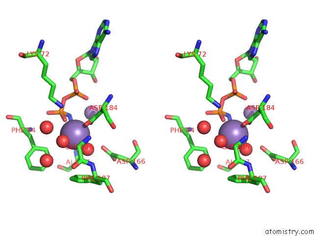
Stereo pair view
Mono view
Stereo pair view
A full contact list of Manganese with other atoms in the Mn binding
site number 2 of A Complex Structure Between the Catalytic and Regulatory Subunit of Protein Kinase A That Represents the Inhibited State within 5.0Å range:
|
Reference:
C.Kim,
C.Y.Cheng,
S.A.Saldanha,
S.S.Taylor.
Pka-I Holoenzyme Structure Reveals A Mechanism For Camp-Dependent Activation. Cell(Cambridge,Mass.) V. 130 1032 2007.
ISSN: ISSN 0092-8674
PubMed: 17889648
DOI: 10.1016/J.CELL.2007.07.018
Page generated: Sat Oct 5 14:59:55 2024
ISSN: ISSN 0092-8674
PubMed: 17889648
DOI: 10.1016/J.CELL.2007.07.018
Last articles
I in 4F4BI in 4E27
I in 4EUU
I in 4EUT
I in 4EBK
I in 4E9O
I in 4DZL
I in 4E91
I in 4DZN
I in 4DZM