Manganese »
PDB 2pal-2qgi »
2pyj »
Manganese in PDB 2pyj: PHI29 Dna Polymerase Complexed with Primer-Template Dna and Incoming Nucleotide Substrates (Ternary Complex)
Enzymatic activity of PHI29 Dna Polymerase Complexed with Primer-Template Dna and Incoming Nucleotide Substrates (Ternary Complex)
All present enzymatic activity of PHI29 Dna Polymerase Complexed with Primer-Template Dna and Incoming Nucleotide Substrates (Ternary Complex):
2.7.7.7;
2.7.7.7;
Protein crystallography data
The structure of PHI29 Dna Polymerase Complexed with Primer-Template Dna and Incoming Nucleotide Substrates (Ternary Complex), PDB code: 2pyj
was solved by
A.J.Berman,
S.Kamtekar,
J.L.Goodman,
J.M.Lazaro,
M.De Vega,
L.Blanco,
M.Salas,
T.A.Steitz,
with X-Ray Crystallography technique. A brief refinement statistics is given in the table below:
| Resolution Low / High (Å) | 41.07 / 2.03 |
| Space group | P 1 21 1 |
| Cell size a, b, c (Å), α, β, γ (°) | 72.835, 114.667, 104.761, 90.00, 94.07, 90.00 |
| R / Rfree (%) | 18.9 / 23.4 |
Other elements in 2pyj:
The structure of PHI29 Dna Polymerase Complexed with Primer-Template Dna and Incoming Nucleotide Substrates (Ternary Complex) also contains other interesting chemical elements:
| Magnesium | (Mg) | 2 atoms |
Manganese Binding Sites:
The binding sites of Manganese atom in the PHI29 Dna Polymerase Complexed with Primer-Template Dna and Incoming Nucleotide Substrates (Ternary Complex)
(pdb code 2pyj). This binding sites where shown within
5.0 Angstroms radius around Manganese atom.
In total 2 binding sites of Manganese where determined in the PHI29 Dna Polymerase Complexed with Primer-Template Dna and Incoming Nucleotide Substrates (Ternary Complex), PDB code: 2pyj:
Jump to Manganese binding site number: 1; 2;
In total 2 binding sites of Manganese where determined in the PHI29 Dna Polymerase Complexed with Primer-Template Dna and Incoming Nucleotide Substrates (Ternary Complex), PDB code: 2pyj:
Jump to Manganese binding site number: 1; 2;
Manganese binding site 1 out of 2 in 2pyj
Go back to
Manganese binding site 1 out
of 2 in the PHI29 Dna Polymerase Complexed with Primer-Template Dna and Incoming Nucleotide Substrates (Ternary Complex)
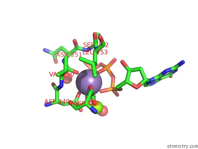
Mono view
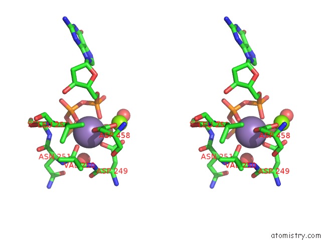
Stereo pair view
Mono view
Stereo pair view
A full contact list of Manganese with other atoms in the Mn binding
site number 1 of PHI29 Dna Polymerase Complexed with Primer-Template Dna and Incoming Nucleotide Substrates (Ternary Complex) within 5.0Å range:
|
Manganese binding site 2 out of 2 in 2pyj
Go back to
Manganese binding site 2 out
of 2 in the PHI29 Dna Polymerase Complexed with Primer-Template Dna and Incoming Nucleotide Substrates (Ternary Complex)
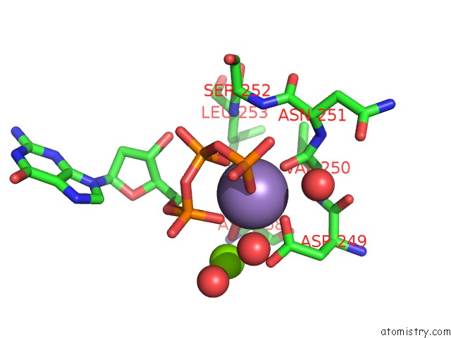
Mono view
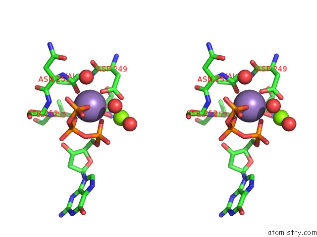
Stereo pair view
Mono view
Stereo pair view
A full contact list of Manganese with other atoms in the Mn binding
site number 2 of PHI29 Dna Polymerase Complexed with Primer-Template Dna and Incoming Nucleotide Substrates (Ternary Complex) within 5.0Å range:
|
Reference:
A.J.Berman,
S.Kamtekar,
J.L.Goodman,
J.M.Lazaro,
M.De Vega,
L.Blanco,
M.Salas,
T.A.Steitz.
Structures of PHI29 Dna Polymerase Complexed with Substrate: the Mechanism of Translocation in B-Family Polymerases Embo J. V. 26 3494 2007.
ISSN: ISSN 0261-4189
PubMed: 17611604
DOI: 10.1038/SJ.EMBOJ.7601780
Page generated: Sat Oct 5 14:57:47 2024
ISSN: ISSN 0261-4189
PubMed: 17611604
DOI: 10.1038/SJ.EMBOJ.7601780
Last articles
K in 2VWJK in 2VXY
K in 2VQW
K in 2VQV
K in 2VQQ
K in 2VQO
K in 2VI5
K in 2VQM
K in 2VQJ
K in 2VPL