Manganese »
PDB 2pal-2qgi »
2phk »
Manganese in PDB 2phk: The Crystal Structure of A Phosphorylase Kinase Peptide Substrate Complex: Kinase Substrate Recognition
Enzymatic activity of The Crystal Structure of A Phosphorylase Kinase Peptide Substrate Complex: Kinase Substrate Recognition
All present enzymatic activity of The Crystal Structure of A Phosphorylase Kinase Peptide Substrate Complex: Kinase Substrate Recognition:
2.7.1.38;
2.7.1.38;
Protein crystallography data
The structure of The Crystal Structure of A Phosphorylase Kinase Peptide Substrate Complex: Kinase Substrate Recognition, PDB code: 2phk
was solved by
E.D.Lowe,
M.E.M.Noble,
V.T.Skamnaki,
N.G.Oikonomakos,
D.J.Owen,
L.N.Johnson,
with X-Ray Crystallography technique. A brief refinement statistics is given in the table below:
| Resolution Low / High (Å) | 25.00 / 2.60 |
| Space group | P 32 2 1 |
| Cell size a, b, c (Å), α, β, γ (°) | 65.300, 65.300, 145.800, 90.00, 90.00, 120.00 |
| R / Rfree (%) | 23.6 / 30 |
Manganese Binding Sites:
The binding sites of Manganese atom in the The Crystal Structure of A Phosphorylase Kinase Peptide Substrate Complex: Kinase Substrate Recognition
(pdb code 2phk). This binding sites where shown within
5.0 Angstroms radius around Manganese atom.
In total 2 binding sites of Manganese where determined in the The Crystal Structure of A Phosphorylase Kinase Peptide Substrate Complex: Kinase Substrate Recognition, PDB code: 2phk:
Jump to Manganese binding site number: 1; 2;
In total 2 binding sites of Manganese where determined in the The Crystal Structure of A Phosphorylase Kinase Peptide Substrate Complex: Kinase Substrate Recognition, PDB code: 2phk:
Jump to Manganese binding site number: 1; 2;
Manganese binding site 1 out of 2 in 2phk
Go back to
Manganese binding site 1 out
of 2 in the The Crystal Structure of A Phosphorylase Kinase Peptide Substrate Complex: Kinase Substrate Recognition
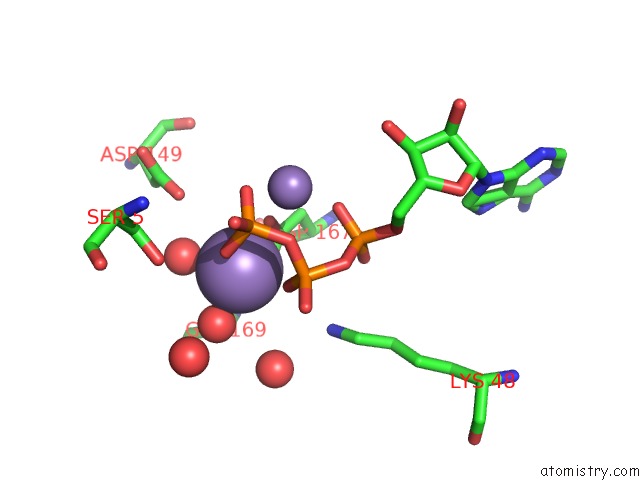
Mono view
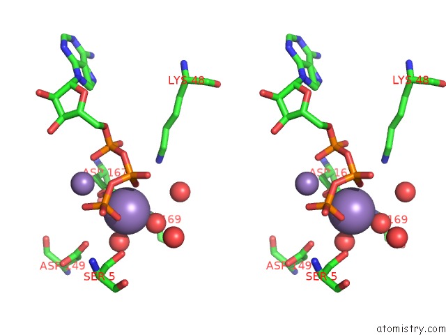
Stereo pair view
Mono view
Stereo pair view
A full contact list of Manganese with other atoms in the Mn binding
site number 1 of The Crystal Structure of A Phosphorylase Kinase Peptide Substrate Complex: Kinase Substrate Recognition within 5.0Å range:
|
Manganese binding site 2 out of 2 in 2phk
Go back to
Manganese binding site 2 out
of 2 in the The Crystal Structure of A Phosphorylase Kinase Peptide Substrate Complex: Kinase Substrate Recognition
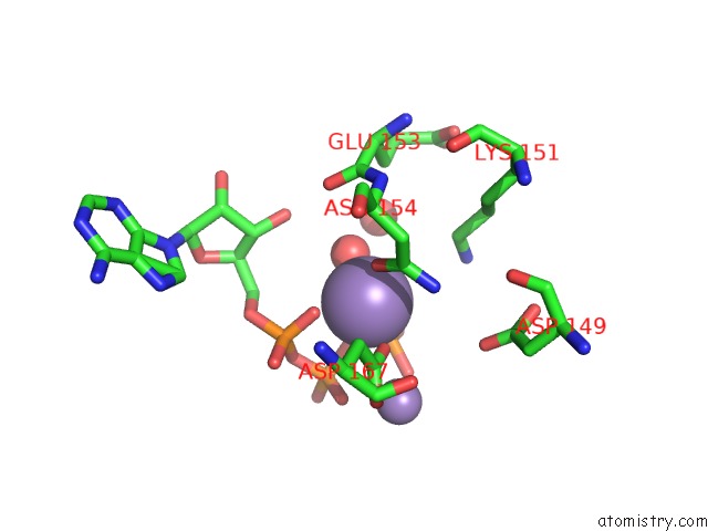
Mono view
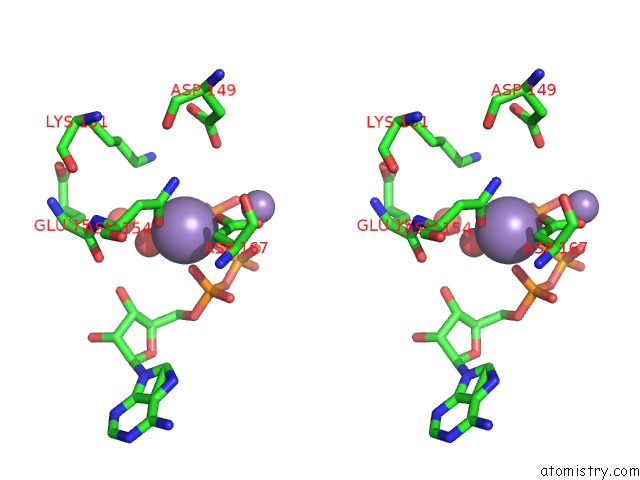
Stereo pair view
Mono view
Stereo pair view
A full contact list of Manganese with other atoms in the Mn binding
site number 2 of The Crystal Structure of A Phosphorylase Kinase Peptide Substrate Complex: Kinase Substrate Recognition within 5.0Å range:
|
Reference:
E.D.Lowe,
M.E.Noble,
V.T.Skamnaki,
N.G.Oikonomakos,
D.J.Owen,
L.N.Johnson.
The Crystal Structure of A Phosphorylase Kinase Peptide Substrate Complex: Kinase Substrate Recognition. Embo J. V. 16 6646 1997.
ISSN: ISSN 0261-4189
PubMed: 9362479
DOI: 10.1093/EMBOJ/16.22.6646
Page generated: Sat Oct 5 14:53:54 2024
ISSN: ISSN 0261-4189
PubMed: 9362479
DOI: 10.1093/EMBOJ/16.22.6646
Last articles
K in 4FXFK in 4G1V
K in 4FXM
K in 4FB0
K in 4FXS
K in 4FO4
K in 4FAW
K in 4FWJ
K in 4FMW
K in 4FLP