Manganese »
PDB 2jck-2nym »
2nym »
Manganese in PDB 2nym: Crystal Structure of Protein Phosphatase 2A (PP2A) with C-Terminus Truncated Catalytic Subunit
Enzymatic activity of Crystal Structure of Protein Phosphatase 2A (PP2A) with C-Terminus Truncated Catalytic Subunit
All present enzymatic activity of Crystal Structure of Protein Phosphatase 2A (PP2A) with C-Terminus Truncated Catalytic Subunit:
3.1.3.16;
3.1.3.16;
Protein crystallography data
The structure of Crystal Structure of Protein Phosphatase 2A (PP2A) with C-Terminus Truncated Catalytic Subunit, PDB code: 2nym
was solved by
Y.Chen,
Y.Xing,
Y.Xu,
Y.Chao,
Z.Lin,
P.D.Jeffrey,
Y.Shi,
with X-Ray Crystallography technique. A brief refinement statistics is given in the table below:
| Resolution Low / High (Å) | 100.00 / 3.60 |
| Space group | P 21 21 21 |
| Cell size a, b, c (Å), α, β, γ (°) | 108.170, 158.860, 270.750, 90.00, 90.00, 90.00 |
| R / Rfree (%) | 26.7 / 33.1 |
Manganese Binding Sites:
The binding sites of Manganese atom in the Crystal Structure of Protein Phosphatase 2A (PP2A) with C-Terminus Truncated Catalytic Subunit
(pdb code 2nym). This binding sites where shown within
5.0 Angstroms radius around Manganese atom.
In total 4 binding sites of Manganese where determined in the Crystal Structure of Protein Phosphatase 2A (PP2A) with C-Terminus Truncated Catalytic Subunit, PDB code: 2nym:
Jump to Manganese binding site number: 1; 2; 3; 4;
In total 4 binding sites of Manganese where determined in the Crystal Structure of Protein Phosphatase 2A (PP2A) with C-Terminus Truncated Catalytic Subunit, PDB code: 2nym:
Jump to Manganese binding site number: 1; 2; 3; 4;
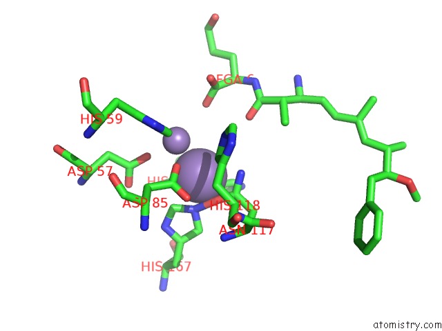
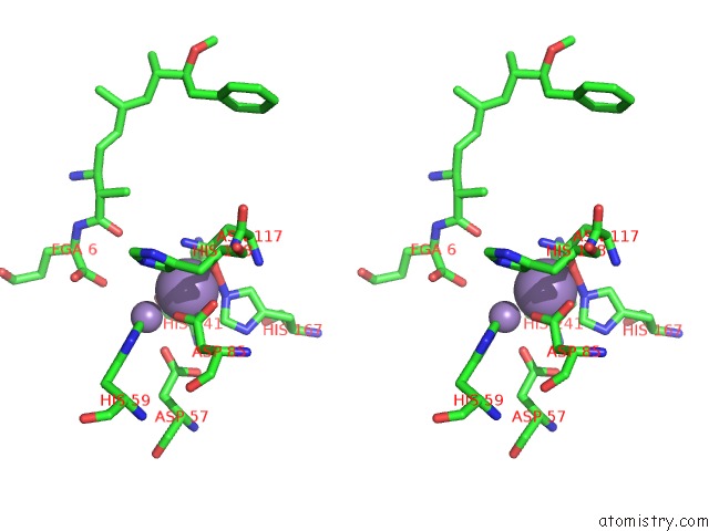
Manganese binding site 1 out of 4 in 2nym
Go back to
Manganese binding site 1 out
of 4 in the Crystal Structure of Protein Phosphatase 2A (PP2A) with C-Terminus Truncated Catalytic Subunit
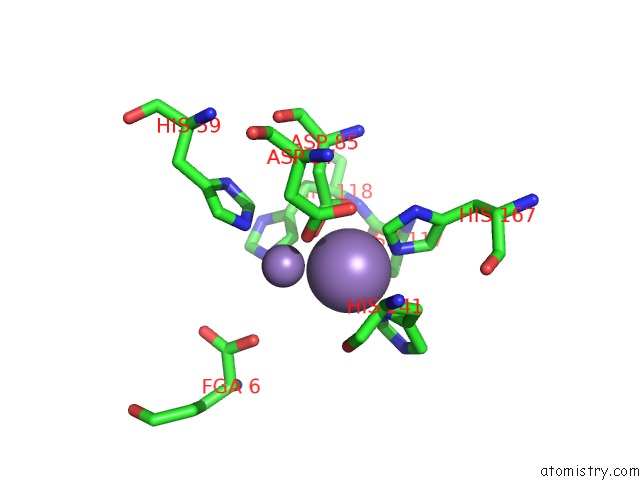
Mono view
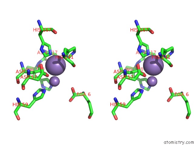
Stereo pair view
Mono view
Stereo pair view
A full contact list of Manganese with other atoms in the Mn binding
site number 1 of Crystal Structure of Protein Phosphatase 2A (PP2A) with C-Terminus Truncated Catalytic Subunit within 5.0Å range:
|
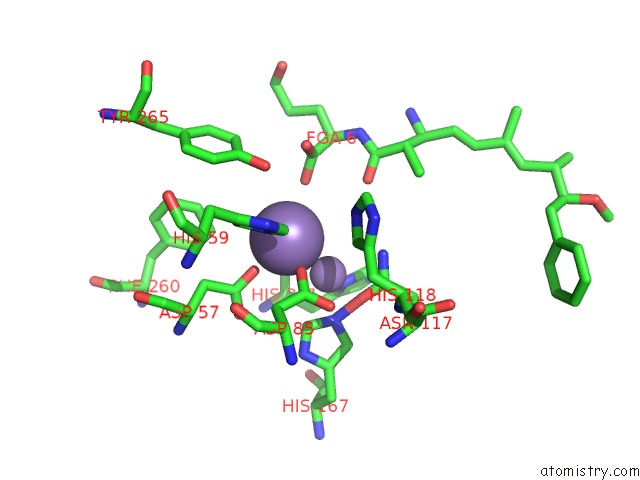
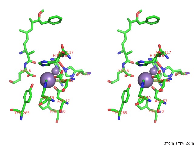
Manganese binding site 2 out of 4 in 2nym
Go back to
Manganese binding site 2 out
of 4 in the Crystal Structure of Protein Phosphatase 2A (PP2A) with C-Terminus Truncated Catalytic Subunit
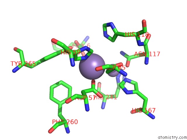
Mono view
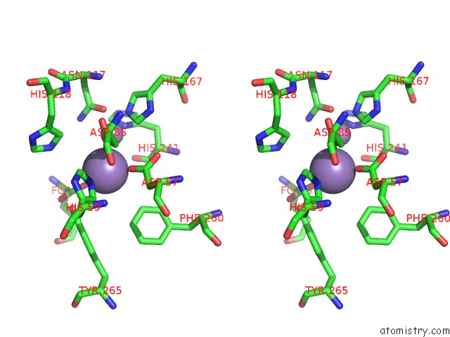
Stereo pair view
Mono view
Stereo pair view
A full contact list of Manganese with other atoms in the Mn binding
site number 2 of Crystal Structure of Protein Phosphatase 2A (PP2A) with C-Terminus Truncated Catalytic Subunit within 5.0Å range:
|
Manganese binding site 3 out of 4 in 2nym
Go back to
Manganese binding site 3 out
of 4 in the Crystal Structure of Protein Phosphatase 2A (PP2A) with C-Terminus Truncated Catalytic Subunit
Mono view
Stereo pair view
Mono view
Stereo pair view
A full contact list of Manganese with other atoms in the Mn binding
site number 3 of Crystal Structure of Protein Phosphatase 2A (PP2A) with C-Terminus Truncated Catalytic Subunit within 5.0Å range:
|
Manganese binding site 4 out of 4 in 2nym
Go back to
Manganese binding site 4 out
of 4 in the Crystal Structure of Protein Phosphatase 2A (PP2A) with C-Terminus Truncated Catalytic Subunit
Mono view
Stereo pair view
Mono view
Stereo pair view
A full contact list of Manganese with other atoms in the Mn binding
site number 4 of Crystal Structure of Protein Phosphatase 2A (PP2A) with C-Terminus Truncated Catalytic Subunit within 5.0Å range:
|
Reference:
Y.Xu,
Y.Xing,
Y.Chen,
Y.Chao,
Z.Lin,
E.Fan,
J.W.Yu,
S.Strack,
P.D.Jeffrey,
Y.Shi.
Structure of the Protein Phosphatase 2A Holoenzyme. Cell(Cambridge,Mass.) V. 127 1239 2006.
ISSN: ISSN 0092-8674
PubMed: 17174897
DOI: 10.1016/J.CELL.2006.11.033
Page generated: Sat Aug 16 10:36:37 2025
ISSN: ISSN 0092-8674
PubMed: 17174897
DOI: 10.1016/J.CELL.2006.11.033
Last articles
Mn in 9LJUMn in 9LJW
Mn in 9LJS
Mn in 9LJR
Mn in 9LJT
Mn in 9LJV
Mg in 9UA2
Mg in 9R96
Mg in 9VM1
Mg in 9P01