Manganese »
PDB 2jck-2nym »
2je7 »
Manganese in PDB 2je7: Crystal Structure of Recombinant Dioclea Guianensis Lectin S131H Complexed with 5-Bromo-4-Chloro-3-Indolyl-A-D-Mannose
Protein crystallography data
The structure of Crystal Structure of Recombinant Dioclea Guianensis Lectin S131H Complexed with 5-Bromo-4-Chloro-3-Indolyl-A-D-Mannose, PDB code: 2je7
was solved by
C.S.Nagano,
L.Sanz,
B.S.Cavada,
J.J.Calvete,
with X-Ray Crystallography technique. A brief refinement statistics is given in the table below:
| Resolution Low / High (Å) | 63.50 / 1.65 |
| Space group | I 2 2 2 |
| Cell size a, b, c (Å), α, β, γ (°) | 65.661, 88.014, 91.536, 90.00, 90.00, 90.00 |
| R / Rfree (%) | 15.4 / 20.1 |
Other elements in 2je7:
The structure of Crystal Structure of Recombinant Dioclea Guianensis Lectin S131H Complexed with 5-Bromo-4-Chloro-3-Indolyl-A-D-Mannose also contains other interesting chemical elements:
| Bromine | (Br) | 1 atom |
| Chlorine | (Cl) | 1 atom |
| Calcium | (Ca) | 1 atom |
Manganese Binding Sites:
The binding sites of Manganese atom in the Crystal Structure of Recombinant Dioclea Guianensis Lectin S131H Complexed with 5-Bromo-4-Chloro-3-Indolyl-A-D-Mannose
(pdb code 2je7). This binding sites where shown within
5.0 Angstroms radius around Manganese atom.
In total only one binding site of Manganese was determined in the Crystal Structure of Recombinant Dioclea Guianensis Lectin S131H Complexed with 5-Bromo-4-Chloro-3-Indolyl-A-D-Mannose, PDB code: 2je7:
In total only one binding site of Manganese was determined in the Crystal Structure of Recombinant Dioclea Guianensis Lectin S131H Complexed with 5-Bromo-4-Chloro-3-Indolyl-A-D-Mannose, PDB code: 2je7:
Manganese binding site 1 out of 1 in 2je7
Go back to
Manganese binding site 1 out
of 1 in the Crystal Structure of Recombinant Dioclea Guianensis Lectin S131H Complexed with 5-Bromo-4-Chloro-3-Indolyl-A-D-Mannose
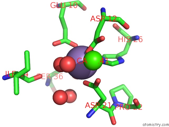
Mono view
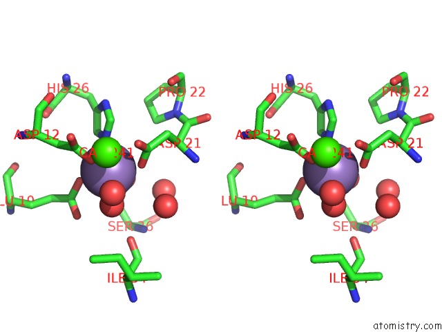
Stereo pair view
Mono view
Stereo pair view
A full contact list of Manganese with other atoms in the Mn binding
site number 1 of Crystal Structure of Recombinant Dioclea Guianensis Lectin S131H Complexed with 5-Bromo-4-Chloro-3-Indolyl-A-D-Mannose within 5.0Å range:
|
Reference:
C.S.Nagano,
J.J.Calvete,
D.Barettino,
A.Perez,
B.S.Cavada,
L.Sanz.
Insights Into the Structural Basis of the pH- Dependent Dimer-Tetramer Equilibrium Through Crystallographic Analysis of Recombinant Diocleinae Lectins. Biochem.J. V. 409 417 2008.
ISSN: ISSN 0264-6021
PubMed: 17937659
DOI: 10.1042/BJ20070942
Page generated: Sat Oct 5 14:33:52 2024
ISSN: ISSN 0264-6021
PubMed: 17937659
DOI: 10.1042/BJ20070942
Last articles
Mg in 6PRVMg in 6PSS
Mg in 6PSR
Mg in 6PSQ
Mg in 6PRY
Mg in 6PRU
Mg in 6PRC
Mg in 6PR5
Mg in 6PQV
Mg in 6PQR